38 label the structures of a skeletal muscle fiber
Art-labeling Activity: The Structure of a Skeletal Muscle Fiber Start studying Art-labeling Activity: The Structure of a Skeletal Muscle Fiber. Learn vocabulary, terms, and more with flashcards, games, and other study tools. Muscle-like fatigue-resistant hydrogels by mechanical training - PNAS May 08, 2019 · Skeletal muscles possess the combinational properties of high fatigue resistance (1,000 J/m 2), high strength (1 MPa), low Young’s modulus (100 kPa), and high water content (70 to 80 wt %), which have not been achieved in synthetic hydrogels.The muscle-like properties are highly desirable for hydrogels’ nascent applications in load-bearing artificial tissues and soft …
en.wikipedia.org › wiki › Central_nervous_systemCentral nervous system - Wikipedia The central nervous system (CNS) is the part of the nervous system consisting primarily of the brain and spinal cord.The CNS is so named because the brain integrates the received information and coordinates and influences the activity of all parts of the bodies of bilaterally symmetric and triploblastic animals—that is, all multicellular animals except sponges and diploblasts.
Label the structures of a skeletal muscle fiber
Solved Hel Label the structures of a skeletal muscle fiber. - Chegg Science; Anatomy and Physiology; Anatomy and Physiology questions and answers; Hel Label the structures of a skeletal muscle fiber. 4 0.1 points eBook Sarcoplasmic reticulum Nucleus Myofibril Openings into T tubules Sarcolemma Mc Graw Hill < Prey 4 of 20 !!! Internal Anatomy of Skeletal Muscle Fibers - GetBodySmart General Anatomy of Skeletal Muscle Fibers An interactive quiz about the general anatomy of skeletal muscle fibers, featuring illustrations-based multiple choice questions. General Organization of the Nervous System Skeletal Muscle Fiber Types - BIO 264 Anatomy & Physiology I Classically, skeletal muscle fibers can be categorized according to their speed of contraction and their resistance to fatigue. These classifications are in the process of being revised, but the basic types include: Slow twitch oxidative (type I) muscle fibers, Fast-twitch oxidative-glycolytic (Type IIA) muscle fibers, and.
Label the structures of a skeletal muscle fiber. Muscle Fibers: Anatomy, Function, and More - Healthline Each muscle fiber contains smaller units made up of repeating thick and thin filaments. This causes the muscle tissue to be striated, or have a striped appearance. Skeletal muscle fibers are... Skeletal Muscle Labeling | Biology Quiz - Quizizz Skeletal Muscle Labeling DRAFT. 22 minutes ago. by ggard. Played 0 times. 0. 9th - 10th grade ... Q. Skeletal Muscle contraction is initiated when the _____ sends a message to the muscle cell. ... Muscle cell. Neuron. Gland. None of the above. Tags: Question 30 . SURVEY . 30 seconds . Report an issue . Q. Each skeletal muscle fiber is ... 11.4 Identify the skeletal muscles and give their origins, … Editor’s note: Replace figure with one that includes all muscles from table for example figure 10.7 from Marieb or 9.8 from Amerman. The orbicularis oris is a circular muscle that moves the lips, and the orbicularis oculi is a circular muscle that closes the eye. The occipitofrontalis muscle elevates the scalp and eyebrows. The muscle has a frontal belly and an occipital belly (near … To label: The structures in the given figure of a skeletal muscle fiber ... Textbook solution for Visual Essentials of Anatomy Physiology 1st Edition Martini Chapter 6.3 Problem 1.4SR. We have step-by-step solutions for your textbooks written by Bartleby experts! To label: The structures in the given figure of a skeletal muscle fiber.
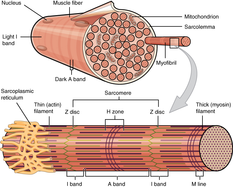
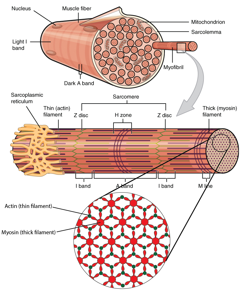
Label the Skeletal Muscle Fiber Quiz - purposegames.com About this Quiz. This is an online quiz called Label the Skeletal Muscle Fiber. There is a printable worksheet available for download here so you can take the quiz with pen and paper.. From the quiz author › doi › 10Muscle-like fatigue-resistant hydrogels by mechanical ... - PNAS May 08, 2019 · The muscle-like properties are highly desirable for hydrogels’ nascent applications in load-bearing artificial tissues and soft devices. Here, we propose a strategy of mechanical training to achieve the aligned nanofibrillar architectures of skeletal muscles in synthetic hydrogels, resulting in the combinational muscle-like properties. Skeletal Muscle Fiber Structure and Function - Open Textbooks for Hong Kong The striated appearance of skeletal muscle tissue is a result of repeating bands of the proteins actin and myosin that occur along the length of myofibrils. Myofibrils are composed of smaller structures called myofilaments. There are two main types of myofilaments: thick filaments and thin filaments. Correctly Label The Following Parts Of A Skeletal Muscle Fiber A skeletal muscle fiber is composed of a plasma membrane and a specialized smooth endoplasmic reticulum. It also contains sarcomeres and calcium ions. In addition to the plasma membrane, a skeletal muscle fiber has numerous myofibrils. During a contraction, the force is transmitted through the tendon to the bone, producing a skeletal movement.
skeletal muscle fiber diagram muscle skeletal anatomy tissue physiology muscular system diagram human muscles structure medical body organization terminology 2000 coding each levels chart. Structure Of Skeletal Muscle Fiber Royalty Free Stock Images - Image . muscle fiber skeletal structure royalty actin. Skeletal Muscle Microanatomy 2 - YouTube www ... en.wikipedia.org › wiki › Skeletal_muscleSkeletal muscle - Wikipedia Many nuclei are needed by the skeletal muscle cell for the large amounts of proteins and enzymes needed to be produced for the cell's normal functioning. A single muscle fiber can contain from hundreds to thousands of nuclei. A muscle fiber for example in the human biceps with a length of 10 cm can have as many as 3000 nuclei. PDF Biology 201: Muscles & Muscle Tissue 1) Label the structures of a ... Source Lesson: Muscle Fiber: Structures, Contraction & Relaxation 4) Label the structures in the diagram below. Some terms may be used more than once. Skeletal Muscle Fiber Structure | The Musculoskeletal System Skeletal Muscle Fiber Structure. Each skeletal muscle fiber is a skeletal muscle cell. These cells are incredibly large, with diameters of up to 100 µm and lengths of up to 30 cm. The plasma membrane of a skeletal muscle fiber is called the sarcolemma.The sarcolemma is the site of action potential conduction, which triggers muscle contraction.
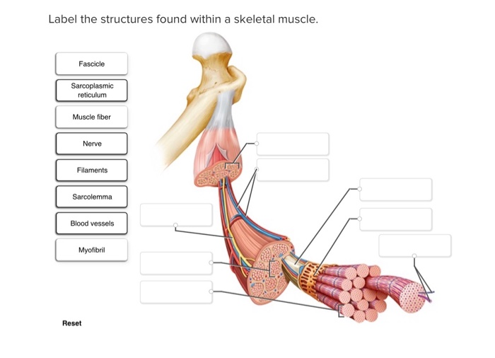
Solved Label the structures found within a skeletal muscle ...
Skeletal muscle - Wikipedia Skeletal muscles (commonly referred to as muscles) are organs of the vertebrate muscular system that are mostly attached by tendons to bones of the skeleton. The muscle cells of skeletal muscles are much longer than in the other types of muscle tissue, and are often known as muscle fibers. The muscle tissue of a skeletal muscle is striated – having a striped …

36 Endomysium Images, Stock Photos & Vectors | Shutterstock
Chapter 9 Homework Flashcards | Quizlet The site where a motor neuron excites a skeletal muscle fiber is called the neuromuscular junction. This activity will test your understanding of the sequence of events that occur at the neuromuscular junction. ... (NMJ): the motor neuron and the skeletal muscle fiber. Label each component with the most appropriate and specific label provided ...

Ultrastructure of a skeletal muscle fiber. (a) Arrangement of ...
Structure of Skeletal Muscle Fibers - Pharmacy 180 Skeletal muscle fibers are the longest types of muscle cells. They contain smaller, rod-shaped myofilaments. The light bands (I bands) are made up of thin filaments of actinsmall, oval-shaped mitochondria and nuclei. Skeletal muscle fibers have multiple nuclei.
Skeletal Muscle Tissue Anatomy and Structure
coursehelponline.comCourse Help Online - Have your academic paper written by a ... All our academic papers are written from scratch. All our clients are privileged to have all their academic papers written from scratch. These papers are also written according to your lecturer’s instructions and thus minimizing any chances of plagiarism.
Structure of Skeletal Muscle – Earth's Lab
› gene › 34793479 - Gene ResultIGF1 insulin like growth factor 1 [ (human)] Skeletal muscle IGF-1 is lower at rest and after resistance exercise in humans with obesity. IGF1 gene is epigenetically activated in preterm infants with intrauterine growth restriction. A high-throughput assay for the quantification of intact Insulin-like Growth Factor I in human serum using online SPE-LC-HRMS.
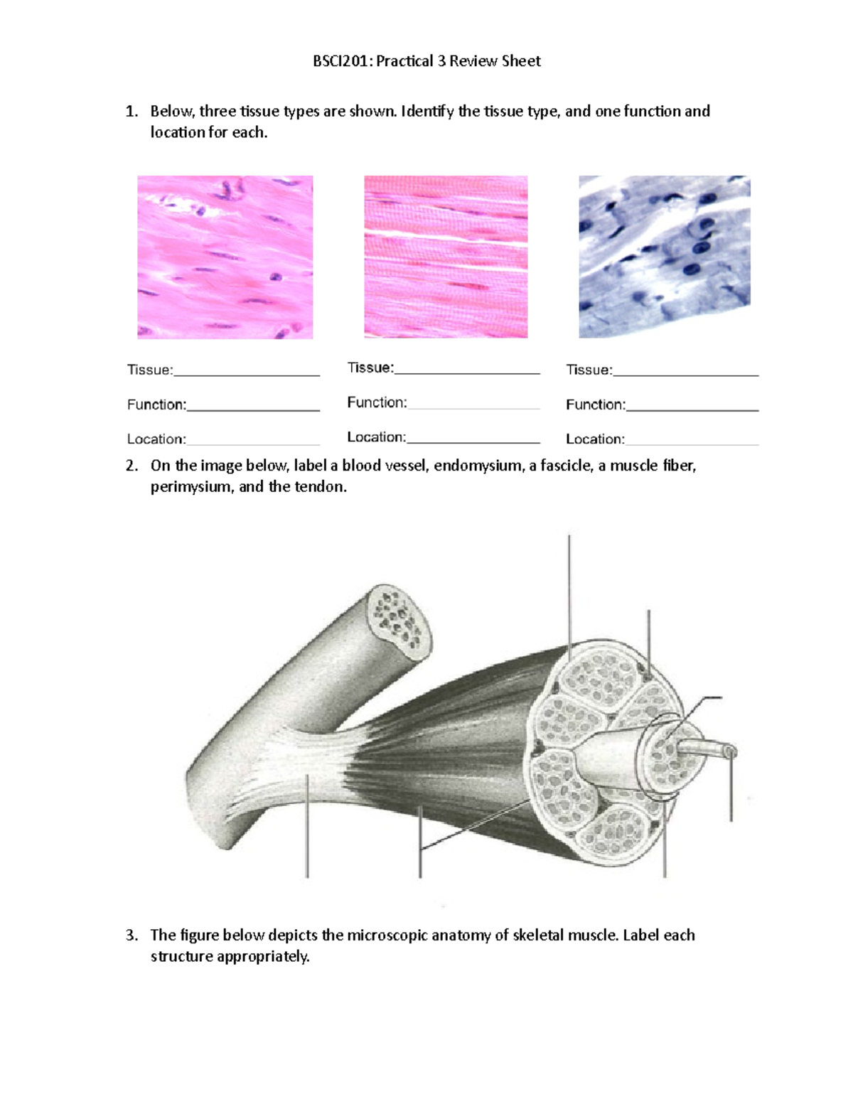
Review of Muscles - 1. Below, three tissue types are shown ...
Solved Muscle Cell Label the structures of a skeletal muscle - Chegg Expert Answer 100% (16 ratings) 1) Sarcolemma 2) myofib … View the full answer Transcribed image text: Muscle Cell Label the structures of a skeletal muscle fiber. Nucleus Myofibril Sarcolemma Sarcoplasmic reticulum Openings into T tubules < Prev 3 of 15 !!! Next > Thinkinys - How to write a boty The Good Cre.

Structure of a Skeletal Muscle Fiber Quiz
Skeletal Muscle Histology Slide Identification and Labeled Diagram From the skeletal muscle histology slide, you might identify the following important structures under the light microscope. Please try to find out these structures from the skeletal muscle slide labeled images. #1. Longitudinal section of skeletal muscle #2. Cross-section of skeletal muscle #3. Skeletal muscle fibers of the longitudinal section #3.
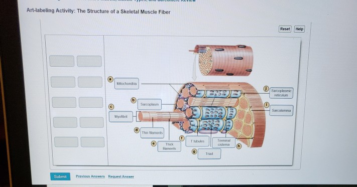
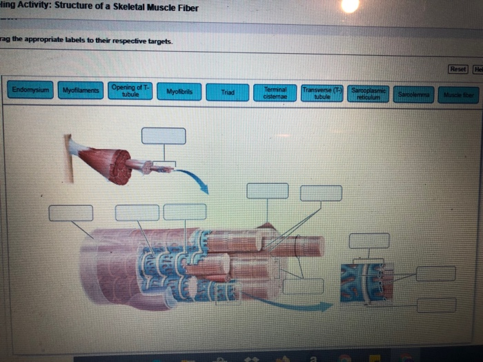
Solved Art-labeling Activity: The Structure of a Skeletal ...
19.4 Cardiac Physiology – Anatomy & Physiology High levels of calcium ions (hypercalcemia) may be implicated in a short QT interval and a widened T wave in the ECG. The QT interval represents the time from the start of depolarization to repolarization of the ventricles, and includes the period of ventricular systole. As in skeletal muscle, increased calcium increases the force of contraction.
Muscles Labeling
Label structure of skeletal muscle Diagram | Quizlet Label structure of skeletal muscle 4.0 5 Reviews STUDY Learn Write Test PLAY Match + − Created by danielaaaa04 Terms in this set (8) myofibrils ... sarcoplasmis reticulum ... sarcolemma ... epimysium ... perimysium ... endomysium ... fascicle ... muscle fiber ... Sets found in the same folder Sarcomere 11 terms MrDNorman TEACHER
STRUCTURE OF SKELETAL MUSCLE
City of Calgary (@cityofcalgary) | Twitter Aug 21, 2008 · Official City of Calgary local government Twitter account. Keep up with City news, services, programs, events and more. Not monitored 24/7.
Solved] Label all the parts of the follow diagrams. | Course Hero
General Anatomy of Skeletal Muscle Fibers - GetBodySmart General Anatomy of Skeletal Muscle Fibers. Start Quiz. Identify and solve gaps in your knowledge using these interactive, spaced repetition-inspired anatomy quizzes. Learn anatomy faster and. remember everything you learn.

11.2 Muscles and Movement | BioNinja
To label: The given structure in the diagram of a skeletal muscle fiber ... Textbook solution for EBK VISUAL ANATOMY & PHYSIOLOGY 3rd Edition Petti Chapter 9.1 Problem 9SR. We have step-by-step solutions for your textbooks written by Bartleby experts! To label: The given structure in the diagram of a skeletal muscle fiber.

Biochemical and structural basis of the passive mechanical ...
Skeletal Muscle Fiber Labeling - Printable About this Worksheet. This is a free printable worksheet in PDF format and holds a printable version of the quiz Skeletal Muscle Fiber Labeling.By printing out this quiz and taking it with pen and paper creates for a good variation to only playing it online.
Skeletal muscle - Wikipedia
quizlet.com › 532891390 › week-6-muscle-physiologyWeek 6: Muscle Physiology Flashcards & Practice Test - Quizlet Sodium channel : a type of voltage-gated ion channel located on the sarcolemma of the muscle fiber. Acetylcholine : neurotransmitter that stimulates skeletal muscle contraction. Acetylcholinesterase : enzyme located in the synaptic cleft that breaks down acetylcholine. Synaptic cleft : the space between the axon terminal and junctional folds.
Solved -ling Activity: Structure of a Skeletal Muscle Fiber ...
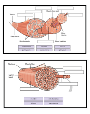
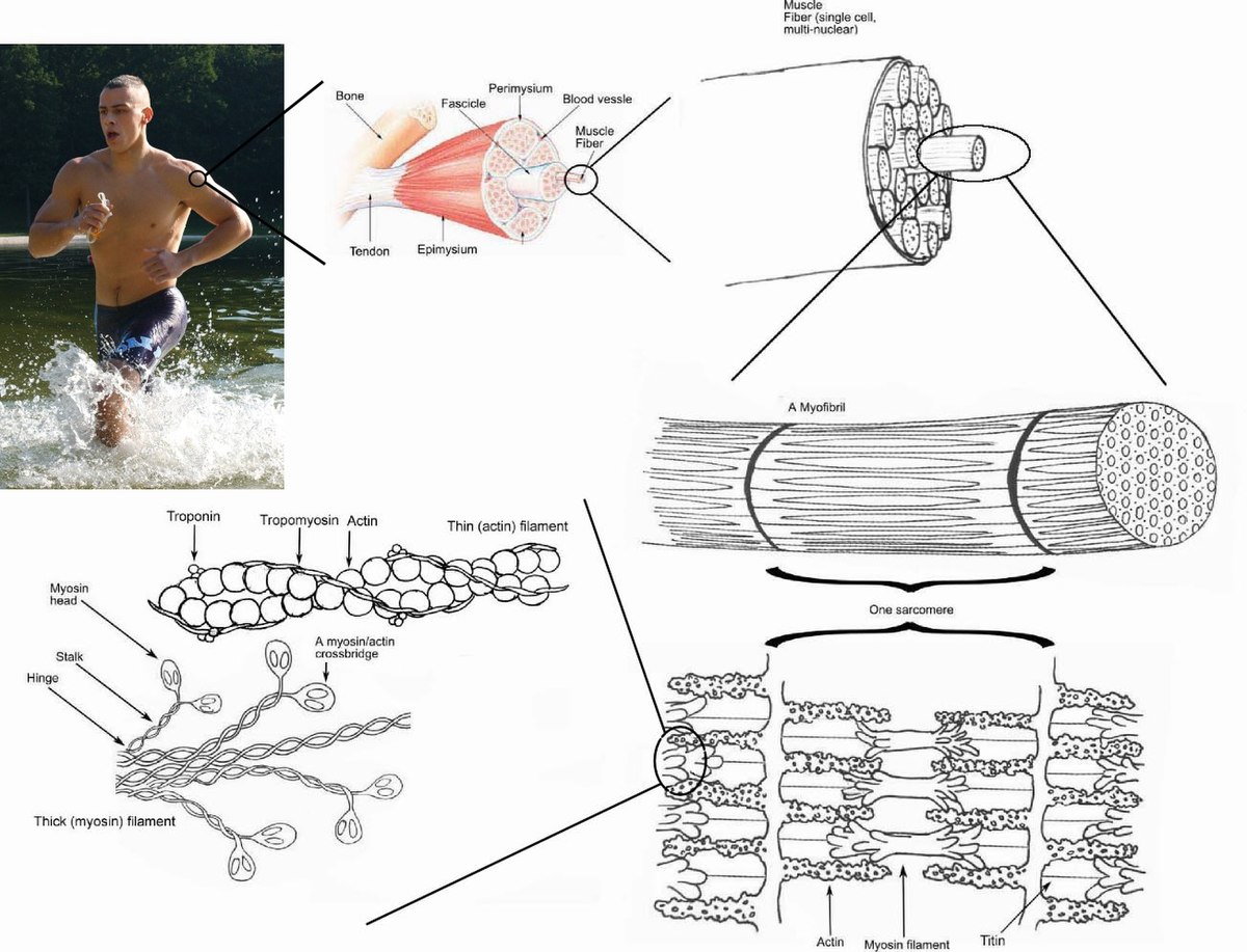
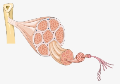
Skeletal Muscle Organization - Cas Skeletal Muscle Cells-Gross and Microscopic Structure. Each skeletal muscle cell, also called a muscle fiber, develops as many embryonic myocytes fused into one long, multi-nucleated skeletal muscle cell. These muscle fibers are bound together into bundles, or fascicles, and are supplied with a rich network of blood vessels and nerves. The ...
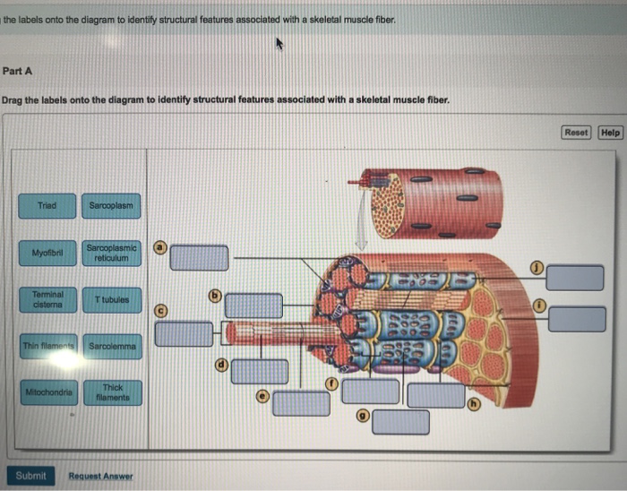
Solved the labels onto the diagram to identify structural ...
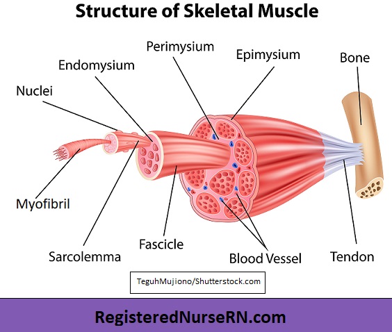
Structure of Skeletal Muscle | SEER Training An individual skeletal muscle may be made up of hundreds, or even thousands, of muscle fibers bundled together and wrapped in a connective tissue covering. Each muscle is surrounded by a connective tissue sheath called the epimysium. Fascia, connective tissue outside the epimysium, surrounds and separates the muscles.

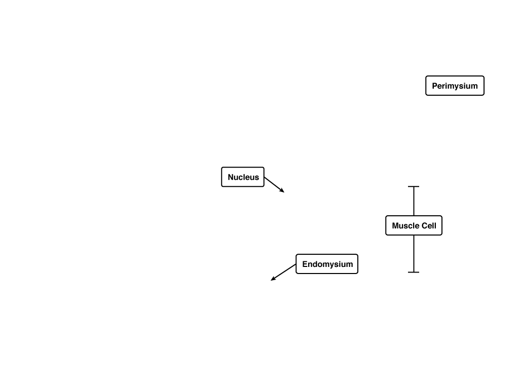
Draw the basic structure of skeletal muscle. Label the ...
3479 - Gene ResultIGF1 insulin like growth factor 1 [ (human)] The protein encoded by this gene is similar to insulin in function and structure and is a member of a family of proteins involved in mediating growth and development. The encoded protein is processed from a precursor, bound by a specific receptor, and secreted. Defects in this gene are a cause of insulin-like growth factor I deficiency.
Skeletal Muscle: Longitudinal Section
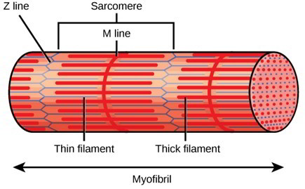
Structure of Skeletal Muscle - CliffsNotes Within a myofibril, actin and myosin filaments are parallel and arranged side by side. The overlapping filaments produce a repeating pattern that gives skeletal muscle its striated appearance. Each repeating unit of the pattern, called a sarcomere, is separated by a border, or Z disc (Z line), to which the actin filaments are attached.

Structures, Superficial - Anatomy and Physiology - Assignment ...
Week 6: Muscle Physiology Flashcards & Practice Test - Quizlet In the Focus Figure, examine the two cells forming the neuromuscular junction (NMJ): the motor neuron and the skeletal muscle fiber. Label each component with the most appropriate and specific label provided. Signals flowing through the neuromuscular junction pass through several structures in a single direction.

Week 6: Muscle Physiology Flashcards | Quizlet
Art labeling Activity The Structure of a Skeletal Muscle Fiber Drag the ... Art labeling Activity The Structure of a Skeletal Muscle Fiber Drag the labels from AA 1. Study Resources. Main Menu; by School; ... Art labeling activity the structure of a skeletal. School No School; Course Title AA 1; Uploaded By JusticeWillpower7289. ... Module 6 Laboratory.pdf. Physiology; Anatomy; 75 pages. Module 6 Laboratory.pdf. SUNY ...
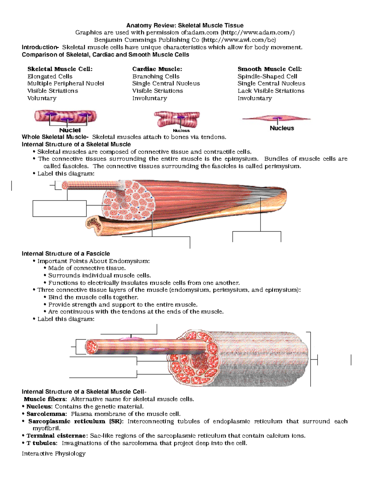
Anatomy Review Skeletal Muscle Tissue - Anatomy Review ...
Skeletal Muscle Fiber | Types, Characteristics & Anatomy - Video ... Structure of Skeletal Muscle Fiber Skeletal muscle fibers are composed of a bundle of thin filaments called myofibrils. Each myofibril is made up of small sections called sarcomeres. Sarcomeres are...
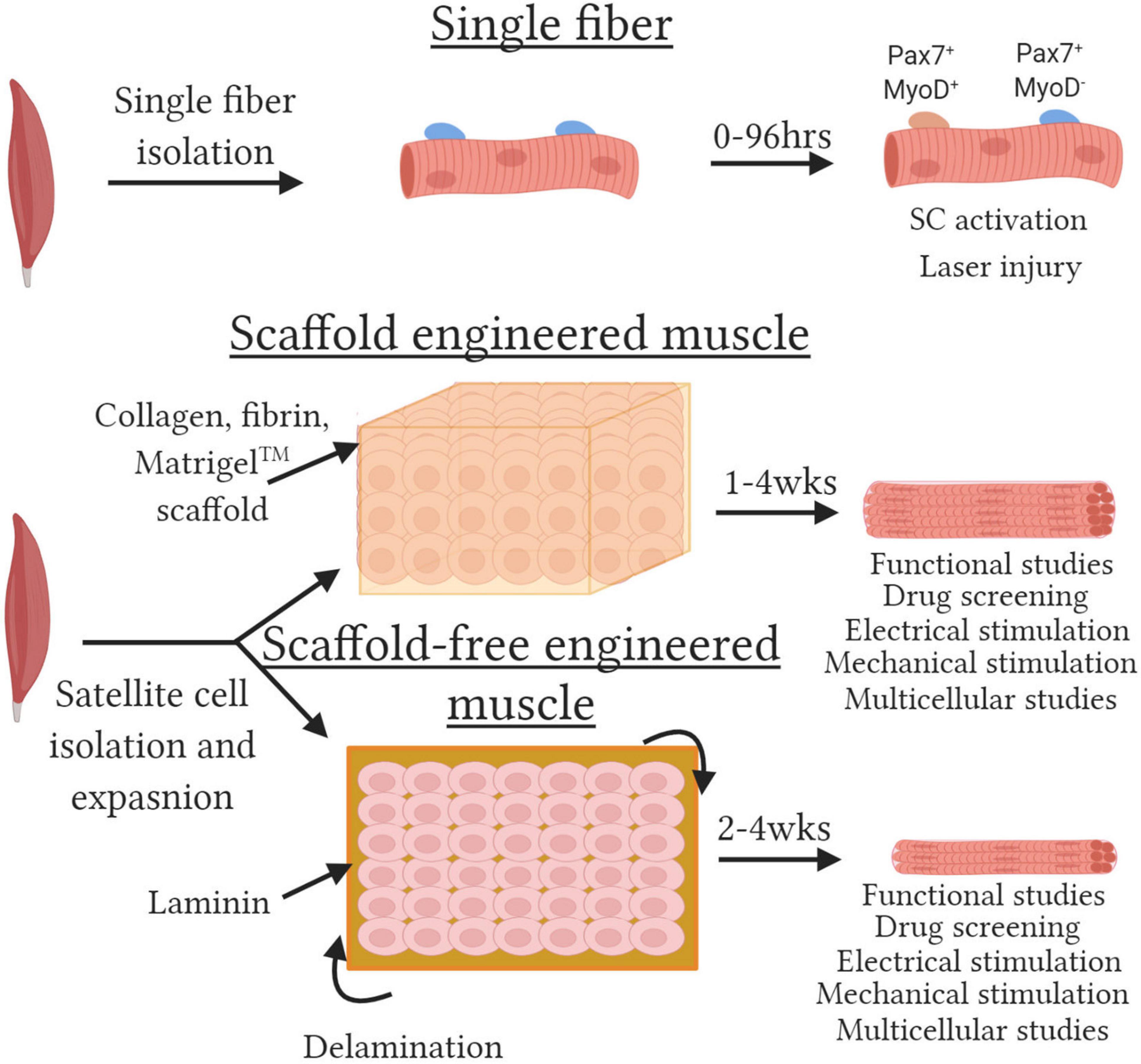
Frontiers | Tissue-Engineered Skeletal Muscle Models to Study ...
open.oregonstate.education › aandp › chapter11.4 Identify the skeletal muscles and give their origins ... This muscle also creates skeletal muscle sphincters at the urethra and anus. Figure 11.4.12 – EDITOR’S NOTE: ADD ISCHIOCOCCYGEUS MUSCLE LABEL TO FIGURE Muscles of the Pelvic Floor: The pelvic floor muscles support the pelvic organs, resist intra-abdominal pressure, and work as sphincters for the urethra, rectum, and vagina.

Scheme of skeletal muscle and associated structures. (Left ...
Course Help Online - Have your academic paper written by a … Professional academic writers. Our global writing staff includes experienced ENL & ESL academic writers in a variety of disciplines. This lets us find the most appropriate writer for …
Muscle Cell Without Labels, HD Png Download - kindpng
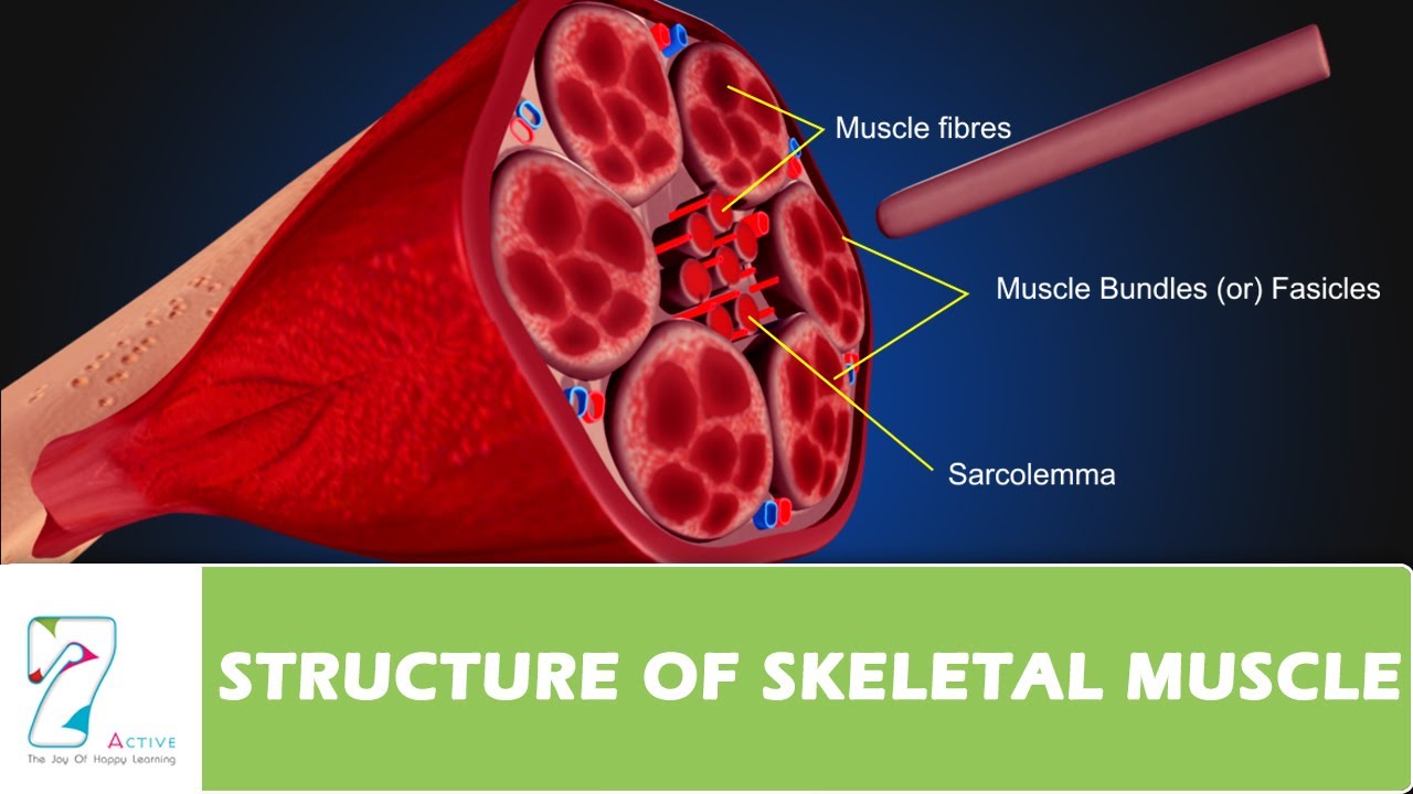
10.2 Skeletal Muscle - Anatomy and Physiology 2e | OpenStax Inside each skeletal muscle, muscle fibers are organized into individual bundles, each called a fascicle, by a middle layer of connective tissue called the perimysium.This fascicular organization is common in muscles of the limbs; it allows the nervous system to trigger a specific movement of a muscle by activating a subset of muscle fibers within a bundle, or fascicle of the muscle.

Diagram of Skeletal Muscle
Skeletal Muscle - Anatomy & Physiology - UH Pressbooks A skeletal muscle fiber is surrounded by a plasma membrane called the sarcolemma, which contains sarcoplasm, the cytoplasm of muscle cells. A muscle fiber is composed of many fibrils, which give the cell its striated appearance. The Sarcomere

Muscle structure – muscle under the microscope — Science ...
Central nervous system - Wikipedia The central nervous system (CNS) is the part of the nervous system consisting primarily of the brain and spinal cord.The CNS is so named because the brain integrates the received information and coordinates and influences the activity of all parts of the bodies of bilaterally symmetric and triploblastic animals—that is, all multicellular animals except sponges and diploblasts.

Lesson Explainer: Structure of Muscles | Nagwa
Skeletal Muscle Fiber Types - BIO 264 Anatomy & Physiology I Classically, skeletal muscle fibers can be categorized according to their speed of contraction and their resistance to fatigue. These classifications are in the process of being revised, but the basic types include: Slow twitch oxidative (type I) muscle fibers, Fast-twitch oxidative-glycolytic (Type IIA) muscle fibers, and.

Label structure of skeletal muscle Diagram | Quizlet
Internal Anatomy of Skeletal Muscle Fibers - GetBodySmart General Anatomy of Skeletal Muscle Fibers An interactive quiz about the general anatomy of skeletal muscle fibers, featuring illustrations-based multiple choice questions. General Organization of the Nervous System
SISTEM OTOT MANUSIA
Solved Hel Label the structures of a skeletal muscle fiber. - Chegg Science; Anatomy and Physiology; Anatomy and Physiology questions and answers; Hel Label the structures of a skeletal muscle fiber. 4 0.1 points eBook Sarcoplasmic reticulum Nucleus Myofibril Openings into T tubules Sarcolemma Mc Graw Hill < Prey 4 of 20 !!!
Skeletal Muscle | Anatomy and Physiology | | Course Hero
OVERVIEW OF MUSCLE TISSUE

STRUCTURE OF A SKELETAL MUSCLE FIBER Diagram | Quizlet

10.2 Skeletal Muscle – Anatomy & Physiology
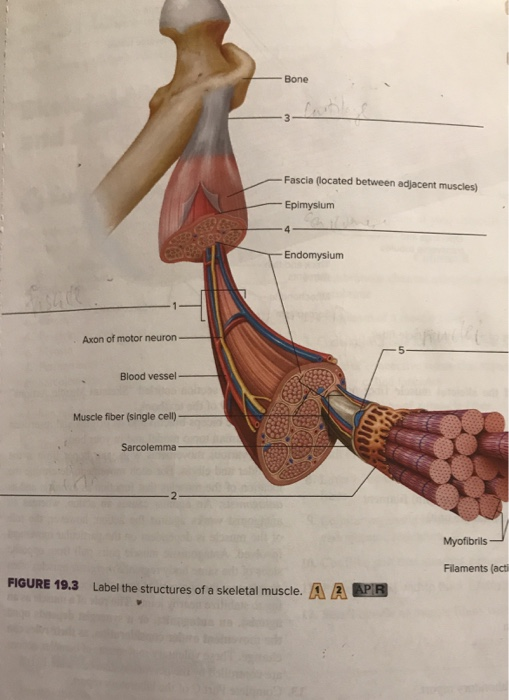
Solved Bone Fascia (located between adjacent muscles) | Chegg.com
Anatomy Review: Skeletal Muscle Tissue
SKELETAL MUSCLE ORGANIZATION

Label the following in a diagram of a skeletal muscle fiber ...

ANSWER THIS NOW!!!! 40 points!!! Drag each label to the ...
Post a Comment for "38 label the structures of a skeletal muscle fiber"