44 diagram for labelling microscope
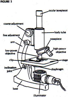
Labelled Diagram of Compound Microscope - Biology Discussion The below mentioned article provides a labelled diagram of compound microscope. Part # 1. The Stand: The stand is made up of a heavy foot which carries a curved inclinable limb or arm bearing the body tube. The foot is generally horse shoe-shaped structure (Fig. 2) which rests on table top or any other surface on which the microscope in kept. Label the microscope — Science Learning Hub Label the microscope Add to collection Use this interactive to identify and label the main parts of a microscope. Drag and drop the text labels onto the microscope diagram. eye piece lens coarse focus adjustment high-power objective diaphragm or iris base fine focus adjustment light source stage Download Exercise Tweet
Label a microscope - Teaching resources - Wordwall Labelling a Microscope Labelled diagram. by Shonprebble. KS3 Y7 Science. Preparing a microscope slide. Match up. by Mmudie. KS3 Biology. Label a plant Year 1 Labelled diagram. by Sciencebowlingpark. KS1 Y1 Science Plants. Parts of a Microscope - Easier Match up. by Msxmdg. KS3 KS4 Science. Microscope quiz Quiz.

Diagram for labelling microscope
Label the Microscope Diagram | Download Scientific Diagram Download scientific diagram | Label the Microscope Diagram from publication: Laboratory Exercises in Microbiology: Discovering the Unseen World through Hands-on Investigation | Microbiology ... Microscope Drawing | How To Draw A Microscope Diagram - YouTube How to draw a microscope diagram. Microscope drawing. Easy and simple step by step tutorial for beginners.Thanks for Watching and Subscribing "Minutes Draw"M... Compound Microscope Parts, Functions, and Labeled Diagram Compound Microscope Parts, Functions, and Labeled Diagram Posted by Fred Koenig on Nov 18th 2020 Compound Microscope Parts, Functions, and Labeled Diagram Parts of a Compound Microscope Each part of the compound microscope serves its own unique function, with each being important to the function of the scope as a whole.
Diagram for labelling microscope. Simple Microscope - Parts, Functions, Diagram and Labelling Simple Microscope - Parts, Functions, Diagram and Labelling A microscope is one of the commonly used equipment in a laboratory setting. A microscope is an optical instrument used to magnify an image of a tiny object; objects that are not visible to the human eyes. Table of Contents The common types of microscopes are: What is a Simple microscope? PDF Label parts of the Microscope Label parts of the Microscope: . Created Date: 20150715115425Z Labeling Microscope Worksheet | Teaching Resources A straightforward worksheet in which students are required to identify the parts of a basic microscope. Tes classic free licence. Report this resource to let us know if it violates our terms and conditions. Our customer service team will review your report and will be in touch. Last updated. 21 November 2014. Microscope labeled diagram - SlideShare Microscope labeled diagram 1. The Microscope Image courtesy of: Microscopehelp.com Basic rules to using the microscope 1. You should always carry a microscope with two hands, one on the arm and the other under the base. 2. You should always start on the lowest power objective lens and should always leave the microscope on the low power lens ...
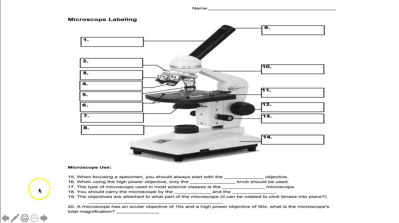
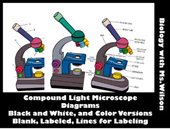
Microscope Labeling - The Biology Corner Students label the parts of the microscope in this photo of a basic laboratory light microscope. Can be used for practice or as a quiz. ... Microscope Labeling . Microscope Use: 15. When focusing a specimen, you should always start with the _____ objective. 16. When using the high power objective, only the _____ knob should be used. 17. The ... Microscope, Microscope Parts, Labeled Diagram, and Functions The Iris Diaphragm is located above the condenser lens and below the microscope stage. The different sized holes in the diaphragm helps to vary the size of the cone and intensity of light that is projected upward into the slide. However, there is no set rule regarding which setting to use for a particular power. Microscope Poster - Diagram with Labels | Teach Starter A poster containing a diagram with labels showing the key parts of a microscope. In Science it is important that students know how to use a variety of tools when conducting scientific experiments and inquiry. This poster focuses on the microscope and highlights its key parts. There are two print options available for this poster: Print on ... Microscope Parts, Function, & Labeled Diagram - slidingmotion Microscope parts labeled diagram gives us all the information about its parts and their position in the microscope. Microscope Parts Labeled Diagram The principle of the Microscope gives you an exact reason to use it. It works on the 3 principles. Magnification Resolving Power Numerical Aperture. Parts of Microscope Head Base Arm Eyepiece Lens
Parts of a Microscope Labeling Activity - Storyboard That Create a poster that labels the parts of a microscope and includes descriptions of what each part does. Click "Start Assignment". Use a landscape poster layout (large or small). Search for a diagram of a microscope. Using arrows and textables label each part of the microscope and describe its function. Copy This Storyboard* More options Parts of the Microscope with Labeling (also Free Printouts) Parts of the Microscope with Labeling (also Free Printouts) A microscope is one of the invaluable tools in the laboratory setting. It is used to observe things that cannot be seen by the naked eye. Table of Contents 1. Eyepiece 2. Body tube/Head 3. Turret/Nose piece 4. Objective lenses 5. Knobs (fine and coarse) 6. Stage and stage clips 7. Aperture Simple Microscope - Diagram (Parts labelled), Principle, Formula and Uses Parts of a Simple Microscope A simple microscope consists of Optical parts Mechanical parts Labeled Diagram of simple microscope parts Optical parts The optical parts of a simple microscope include Lens Mirror Eyepiece Lens A simple microscope uses biconvex lens to magnify the image of a specimen under focus. Microscope Labeling - The Biology Corner 1) Start with scanning (the shortest objective) and only use the COARSE knob . Once it is focused… 2) Switch to low power (medium) and only use the COARSE knob . You may need to recenter your slide. Once it is focused.. 3) Switch to high power (long objective).
2. label the part of the microscope by writing the names of ...
Microscope Types (with labeled diagrams) and Functions Phase-contrast microscope labeled diagram Phase-contrast microscope functions: Its applications areas include In cases where the specimen is colorless and is very tiny In biology to conduct cellular level examination of microorganisms that can't be visualized using the bright field microscopy Interference Microscope

Compound Microscope Parts, Diagram Definition, Application ...
Labelling a Microscope Diagram | Quizlet A diaphragm on a microscope is the piece that enables the user to adjust the amount of light that is focused under the specimen being observed Light Source A light microscope uses focused light and lenses to magnify a specimen, usually a cell.
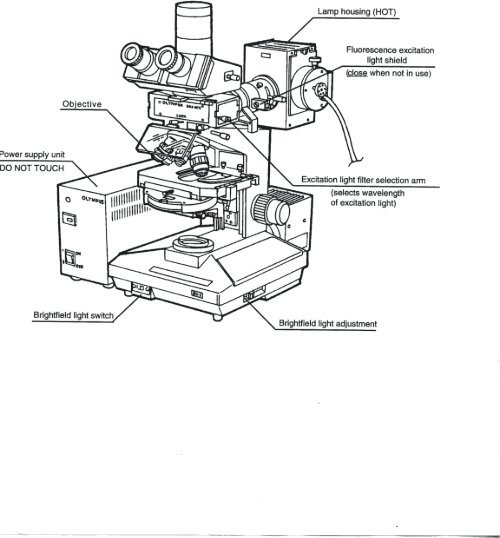
Olympus Research Microscope Diagram - web.biosci.utexas.edu
Compound Microscope Parts - Labeled Diagram and their Functions - Rs ... Labeled diagram of a compound microscope Major structural parts of a compound microscope There are three major structural parts of a compound microscope. The head includes the upper part of the microscope, which houses the most critical optical components, and the eyepiece tube of the microscope.

Microscope Label diagram Diagram | Quizlet
Microscope Parts and Functions Microscope Parts and Functions With Labeled Diagram and Functions How does a Compound Microscope Work?. Before exploring microscope parts and functions, you should probably understand that the compound light microscope is more complicated than just a microscope with more than one lens.. First, the purpose of a microscope is to magnify a small object or to magnify the fine details of a larger ...
Glossary of terms used in microscopy – Quekett Microscopical Club
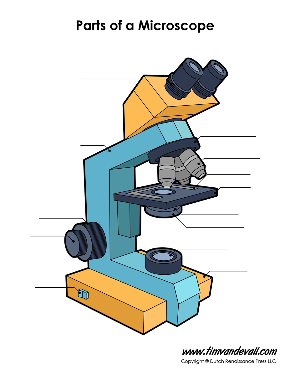
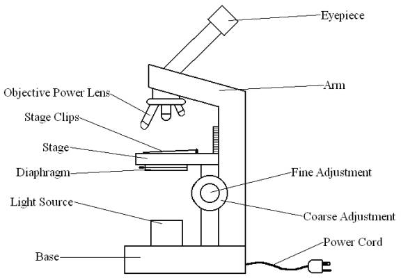
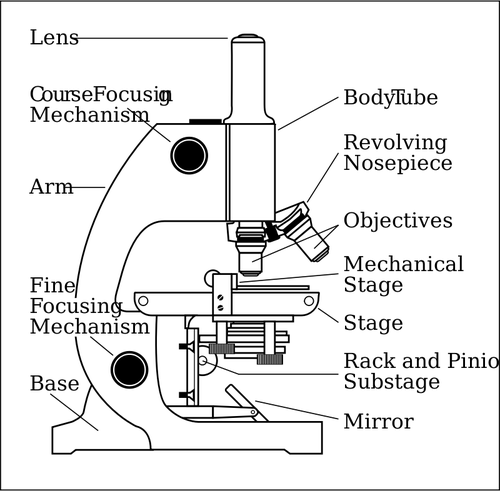
Label Microscope Diagram - EnchantedLearning.com arm - this attaches the eyepiece and body tube to the base. base - this supports the microscope. body tube - the tube that supports the eyepiece. coarse focus adjustment - a knob that makes large adjustments to the focus. diaphragm - an adjustable opening under the stage, allowing different amounts of light onto the stage.
Microscope Labeling
Labelling a Microscope - Labelled diagram - Wordwall Labelling a Microscope. Share Share by Shonprebble. KS3 Y7 Science. Like. Edit Content. Embed. More. Leaderboard. Show more Show less . This leaderboard is currently private. Click Share to make it public. This leaderboard has been disabled by the resource owner. This leaderboard is disabled as your options are different to the resource owner. ...

Diagram of Compound Microscope
Labeling the Parts of the Microscope Labeling the Parts of the Microscope This activity has been designed for use in homes and schools. Each microscope layout (both blank and the version with answers) are available as PDF downloads. You can view a more in-depth review of each part of the microscope here. Download the Label the Parts of the Microscope PDF printable version here.

Microscope parts 3D learning for Android - APK Download
Microscope Labeling Diagram | Quizlet Unit 2 Lesson 5 - Punnett Squares and Pedigrees. 4 terms. PGFry210. Unit 2 Lesson 4 - Heredity. 9 terms. PGFry210. Upgrade to remove ads. Only $2.99/month.
Label Microscope Parts - ClipArt Best
Labeling the Parts of the Microscope | Microscope activity, Science ... Jan 13, 2016 - Free worksheets for labeling parts of the microscope including a worksheet that is blank and one with answers. Pinterest. Today. Explore. ... Print a microscope diagram, microscope worksheet, or practice microscope quiz in order to learn all the parts of a microscope. CCabreza. Biology.
Microscope Diagram To Label - ClipArt Best
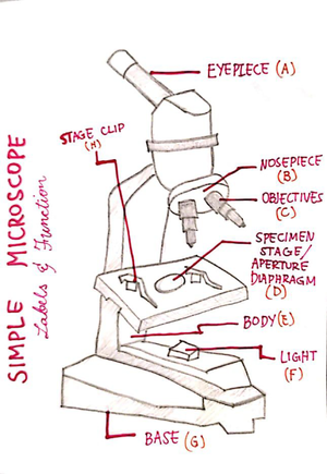
Parts of a microscope with functions and labeled diagram Figure: Diagram of parts of a microscope There are three structural parts of the microscope i.e. head, base, and arm. Head - This is also known as the body. It carries the optical parts in the upper part of the microscope. Base - It acts as microscopes support. It also carries microscopic illuminators.

Microscope Labeling Diagram | Quizlet
Compound Microscope Parts, Functions, and Labeled Diagram Compound Microscope Parts, Functions, and Labeled Diagram Posted by Fred Koenig on Nov 18th 2020 Compound Microscope Parts, Functions, and Labeled Diagram Parts of a Compound Microscope Each part of the compound microscope serves its own unique function, with each being important to the function of the scope as a whole.
Label microscope - Teaching resources
Microscope Drawing | How To Draw A Microscope Diagram - YouTube How to draw a microscope diagram. Microscope drawing. Easy and simple step by step tutorial for beginners.Thanks for Watching and Subscribing "Minutes Draw"M...

✓ microscope view free vector eps, cdr, ai, svg vector ...
Label the Microscope Diagram | Download Scientific Diagram Download scientific diagram | Label the Microscope Diagram from publication: Laboratory Exercises in Microbiology: Discovering the Unseen World through Hands-on Investigation | Microbiology ...

Microscope Mikroskop Monoculer Yazumi XSP-12 + Lampu (Pembesaran 500x) - SINHER/JUNGSON di King Medical | Tokopedia

Microscope Diagram Labeled, Unlabeled and Blank | Parts of a ...

Label a Microscope Worksheet by NC Middle School Resources | TpT
Microscope- Simple-AND Compound-WITH- Label - BS in Education ...

Simple Microscope - Diagram (Parts labelled), Principle ...

Diagrams of Microscope label - YouTube
Label The Parts Of A Compound Microscope Teaching Resources | TpT

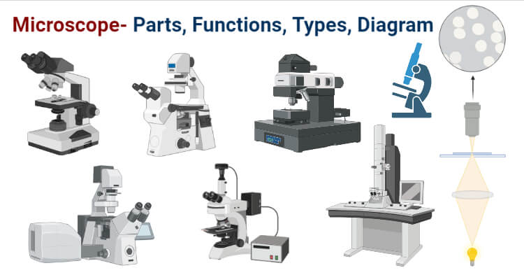
Types of Microscopes: Definition, Working Principle, Diagram ...

Parts of a Microscope with Their Functions • Microbe Online

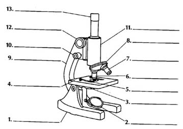
Label the diagram of the microscope with the following parts ...
Boreal Standard Compound Microscope Promotion
Microscope Diagram Labeled, Unlabeled and Blank | Parts of a ...

Diagram Carl Zeiss Mikroskop Optik, Diagram Lembar Kerja ...

Compound Microscope Parts – Labeled Diagram and their ...

Microscope Basics. - ppt download

Parts of a microscope labeling functions worksheet science ...
Microscope- Definition, Parts, Functions, Types, Diagram, Uses
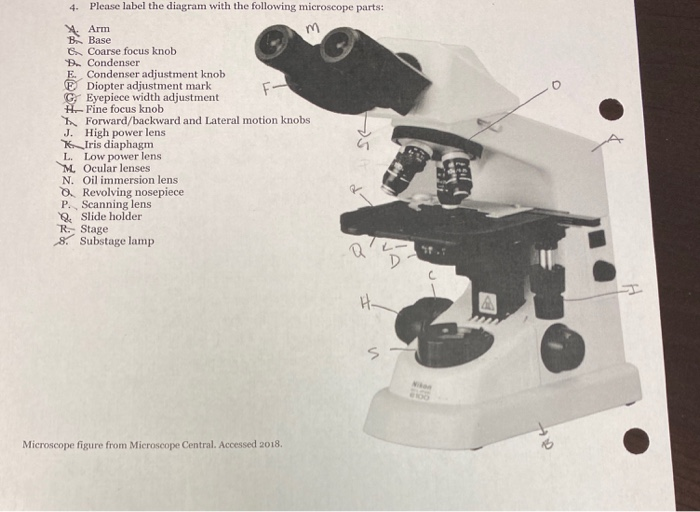
Solved 4. Please label the diagram with the following | Chegg.com

How to Draw a Microscope and Label Its Parts

Compound Microscope Parts – Labeled Diagram and their ...
Compound Microscope Parts, Functions, and Labeled Diagram ...
Label a microscope - Teaching resources
Free Microscope Drawing, Download Free Microscope Drawing png ...

Microscope Parts and Functions
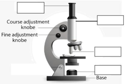
SOLVED:'1. Label the parts of microscope. Course adjustment ...
Parts of the Microscope with Labeling (also Free Printouts ...

Simple Microscope - Parts, Functions, Diagram and Labelling ...
Free Microscope Drawing, Download Free Microscope Drawing png ...
Mikroskop sisi vektor menggambar dengan bagian-bagian yang ...

Microscope Diagram - Label Diagram | Quizlet

Label Microscope Diagram - EnchantedLearning.com
Post a Comment for "44 diagram for labelling microscope"