44 label microscope diagram
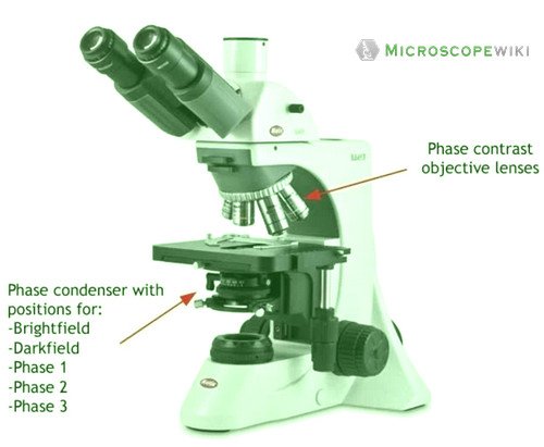
Bright-field microscope (Compound light microscope) - Diagram (Parts ... Bright-field microscope parts (Labeled Diagram) Ocular Lens This microscope has two eye lenses or ocular lens on the top of the microscope that are used to focus the image from the objective lens. It is from these lenses that we see the magnified image of the specimen. Objective Lens Microscope Diagram Worksheet - The Microscope Create A Labelled Diagram ... Using the terms listed below, label the microscope diagram. When you can identify a part of the microscope place the . There is a printable worksheet available for download here so you can take the . Use the words from this word list to identify the parts of the microscope. Use the following terms to correctly label the microscope: Used to ...
rsscience.com › stereo-microscopeParts of Stereo Microscope (Dissecting microscope) – labeled ... Labeled part diagram of a stereo microscope Major structural parts of a stereo microscope. There are three major structural parts of a stereo microscope. The viewing Head includes the upper part of the microscope, which houses the most critical optical components, including the eyepiece, objective lens, and light source of the microscope.

Label microscope diagram
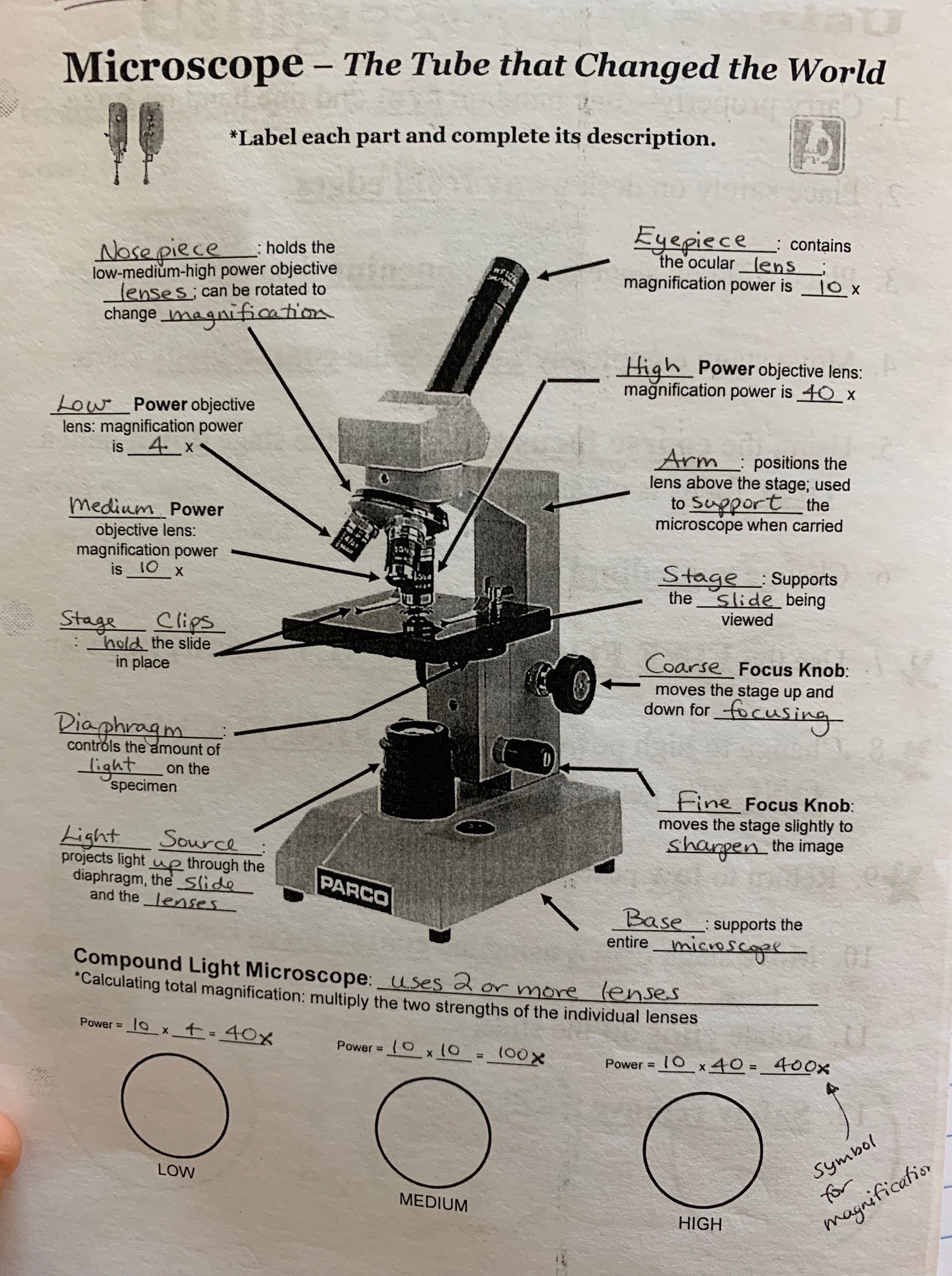
Electron Microscope-Definition, Principle, Types, Uses, Labeled Diagram Parts of an Electron Microscope The electron microscope is placed vertically and has the shape of a tall vacuum column. It consists of the following elements: 1. Electron gun A heated tungsten filament that produces electrons makes up the electron cannon. 2. Electromagnetic lenses The condenser lens directs the electron beam to the specimen. Welcome to Virtual Urchin - University of Washington microscope measurement. microscope compare. specimen compare. development & embryology. fertilization lab. embryogenesis to hatching. analyzing gene function. ecology & environment. our acidifying ocean. predator & prey. surfing to settlement. basic biology. urchin anatomy. about us. teacher resources. useful links . Select Language: Welcome to the new … Label The Parts Of A Microscope Worksheet Answers You can use the word bank below to fill in the blanks or cut. Label the parts of a microscope worksheet answers. Students label the parts of the microscope in this photo of a basic laboratory light microscope. Files include a link to editable doc so you can rewrite a. Power 10 x 4 40 Power 10 x 10 100 Power 10 x 40 400 What happens as the power ...
Label microscope diagram. microscope | Types, Parts, History, Diagram, & Facts microscope, instrument that produces enlarged images of small objects, allowing the observer an exceedingly close view of minute structures at a scale convenient for examination and analysis. Although optical microscopes are the subject of this article, an image may also be enlarged by many other wave forms, including acoustic, X-ray, or electron beam, and be received by direct or digital ... Microscope Diagram - cell division of e coli with continuous media flow ... Microscope Diagram - 15 images - give a well labelled diagram of compound microscope using of typical, bio tem biological transmission electron microscope university, labelled microscope diagram gcse micropedia, a compound microscope diagram micropedia, pE-300white | LED Microscope Illuminator - CoolLED CoolLED Ltd supply LED Microscope Illuminator such as pE-300white for distributors throughout the world. Call +44 (0)1264 323040(UK) or 1.800.877.0128(USA). researchtweet.com › microscope-parts-labeledMicroscope, Microscope Parts, Labeled Diagram, and Functions Jan 19, 2022 · The liquid sample comes next. To minimise evaporation and protect the microscope lens from sample exposure, a small square of clear glass or plastic (a coverslip) is placed on top of the liquid. 1. Collect a clean microscope slide and a coverslip (a thin piece of plastic covering). Fill the centre of the microscope slide with a drop or two of ...
Microscope Types (with labeled diagrams) and Functions Simple microscope labeled diagram Simple microscope functions It is used in industrial applications like: Watchmakers to assemble watches Cloth industry to count the number of threads or fibers in a cloth Jewelers to examine the finer parts of jewelry Miniature artists to examine and build their work Also used to inspect finer details on products Simple Microscope - Parts, Functions, Diagram and Labelling Simple Microscope - Parts, Functions, Diagram and Labelling By Editorial Team March 7, 2022 A microscope is one of the commonly used equipment in a laboratory setting. A microscope is an optical instrument used to magnify an image of a tiny object; objects that are not visible to the human eyes. Table of Contents Brightfield Microscope (Compound Light Microscope)- Definition ... Parts of a microscope with functions and labeled diagram; 22 Types of Spectroscopy with Definition, Principle, Steps, Uses ... Functions, Labeled Diagram; Disadvantages of Brightfield microscope. The aperture diaphragm may cause great contrast which may distort the outcome of the image, therefore iris diaphragm is preferred. Sperm Under Microscope with Labeled Diagram - AnatomyLearner Sperm Under Microscope 400X Labeled Diagram Before that, you may also read the below-mentioned article to get a full idea of the structure of seminiferous tubules - Histological features of the seminiferous tubules with the labeled diagram Okay, first, let's see the different histological features of the seminiferous tubules of an animal.
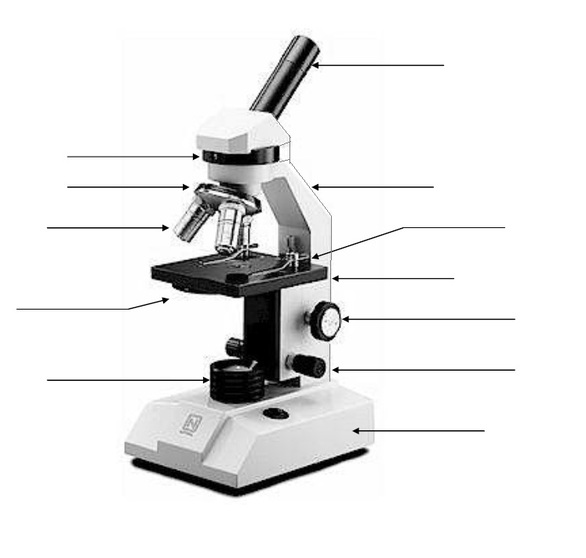
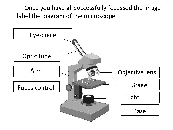
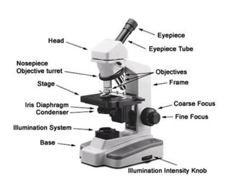
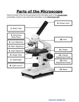
Parts of the Microscope with Labeling (also Free Printouts) Parts of the Microscope with Labeling (also Free Printouts) By Editorial Team March 7, 2022 A microscope is one of the invaluable tools in the laboratory setting. It is used to observe things that cannot be seen by the naked eye. Table of Contents 1. Eyepiece 2. Body tube/Head 3. Turret/Nose piece 4. Objective lenses 5. Knobs (fine and coarse) 6. Inverted Microscope- Definition, Principle, Parts, Labeled Diagram ... Uses of the Inverted Microscope. It is used in diagnostics fungal cultures, for example, detection of Phytophthora spp in cultures. Used for diagnosis of nematology extraction specimens to observe nematodes such as Vermiform nematodes. Used to observe living microbial cells found at the bottom of lab vessels such as tissue culture flasks and ... Light Microscope- Definition, Principle, Types, Parts, Labeled Diagram ... Parts of a microscope with functions and labeled diagram 22 Types of Spectroscopy with Definition, Principle, Steps, Uses History of Microbiology and Contributors in Microbiology Microbiology of extreme environments (Types and Examples) Dark-Field Light Microscope Compound Microscope - Diagram (Parts labelled), Principle and Uses What are the 13 parts of a microscope? 1. Eyepiece 2. Eyepiece Tube 3. Objective Lens 4. Stage 5. Stage Clips 6. Nosepiece 7. Fine and Coarse Focus knobs 8. Illuminator 9. Aperture 10. Iris Diaphragm 11. Condenser 12. Condenser Focus Knob 13. The Rack stop Q 5. What are the 11 parts of a compound microscope?
Produk Microscope | UD Berkah Abadi
Simple Squamous Epithelium under a Microscope with a Labeled Diagram ... From the lung parenchyma labeled diagram, you might identify the following structures - Simple squamous epithelium lining of the lung alveoli (within the parenchyma), A connective tissue basement membrane beneath the simple squamous epithelium lining, The lumen of the lung alveoli, and The cytoplasm of the simple squamous epithelium cells.

This is a common compound microscope Label its parts class 11 ...
microbenotes.com › parts-of-a-microscopeParts of a microscope with functions and labeled diagram Apr 19, 2022 · Figure: Diagram of parts of a microscope. There are three structural parts of the microscope i.e. head, base, and arm. Head – This is also known as the body. It carries the optical parts in the upper part of the microscope. Base – It acts as microscopes support. It also carries microscopic illuminators.
Labeled Parts Of A Microscope - ClipArt Best
Microscope: Types of Microscope, Parts, Uses, Diagram - Embibe There microscope anatomy includes three structural parts, i.e. head, base, and arm. Head - This is also known as the body; it carries the optical parts in the upper part of the microscope.. Base - It acts as microscopes support.It also carries microscopic illuminators. Arms - The microscope arm connects the base and the head and the eyepiece tube to the microscope base.

Microscope Diagram Labeled, Unlabeled and Blank | Parts of a ...
Electron Microscope Principle, Uses, Types and Images (Labeled Diagram ... Electron Microscope Principle, Uses, Types and Images (Labeled Diagram), Price Electron Microscope By Editorial Team Last updated on February 2, 2022 The advances in technology have enabled the development of powerful microscopes to view the samples at a nanometer level and thus were born the electron microscopes.
Microscope Using a microscope I have developed my
Microscope Parts, Function, & Labeled Diagram - slidingmotion Microscope parts labeled diagram gives us all the information about its parts and their position in the microscope. Microscope Parts Labeled Diagram The principle of the Microscope gives you an exact reason to use it. It works on the 3 principles. Magnification Resolving Power Numerical Aperture. Parts of Microscope Head Base Arm Eyepiece Lens

Label Microscope Diagram | Microscope parts, Microscope ...
en.wikipedia.org › wiki › Fluorescence_microscopeFluorescence microscope - Wikipedia A fluorescence microscope is an optical microscope ... design shown in the diagram. ... binding of an antibody to its antigen in order to label specific proteins or ...
Monday 10/19/15 AIM: how do the parts of the compound light ...
Blood Histology Slides with Description and Labeled Diagram The blood is a specialized connective tissue that is fluid and circulates through the vascular channel. In the blood histology slide, you will find different types of cells with their specific features. This might be a short article where I will show you all the cells from the blood microscope slide with a labeled diagram and actual pictures.

Parts of Stereo Microscope (Dissecting microscope) – labeled ...
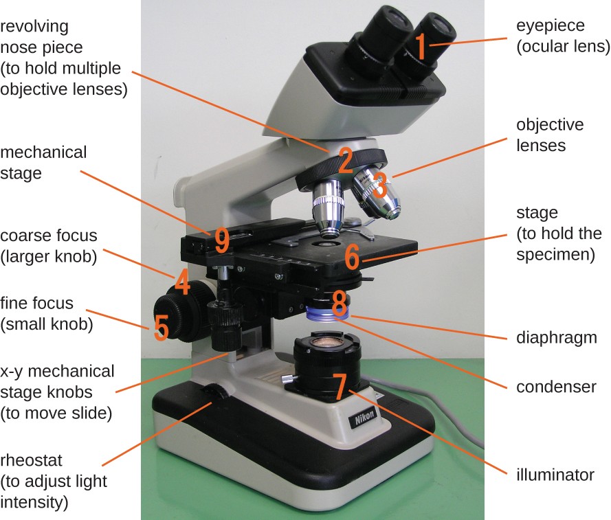
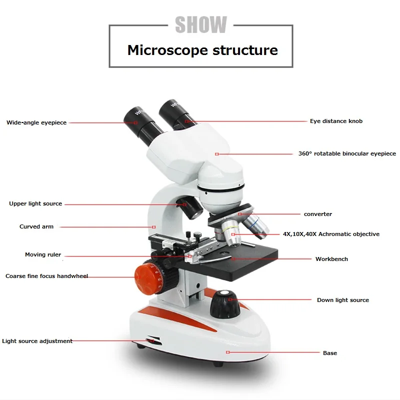
Binocular Microscope Anatomy - Parts and Functions with a Labeled Diagram Now, I will discuss the details anatomy of the light compound microscope with the labeled diagram. Why it is called binocular: because it has two ocular lenses or an eyepiece on the head that attaches to the objective lens, this ocular lens magnifies the image produced by the objective lens. Binocular microscope parts and functions

File:Labelledmicroscope.gif - Wikimedia Commons
Parts of Stereo Microscope (Dissecting microscope) – labeled diagram ... Unlike a compound microscope that offers a flat image, stereo microscopes give the viewer a 3-dimensional image that you can see the texture of a larger specimen. [In this image] Examples of Stereo & Dissecting microscopes. Major microscope brands (Zeiss, Olympus, Nikon, Amscope, Omano, Leica …) all produce stereomicroscopes.
(159).jpg)
Microscope Quiz: How Much You Know About Microscope Parts And ...
Ternary Phase Diagram - an overview | ScienceDirect Topics Ternary phase diagrams are used to represent all possible mixtures of three solvents [1]; they are described in Chapter 3.Here, we shall indicate how they should be used to minimize the solvent consumption. Figure 2.1 (top) shows the methanol–chloroform–water ternary phase diagram with the tie-lines in the biphasic domain. Five particular compositions are shown in the …
microscope parts and functions - Quizizz
Microscopy- History, Classification, Terms, Diagram - The Biology Notes History of Microscope. In the 1 st Century AD, the Romans invented the glass and used them to magnify objects. In the early 14 th Century AD, eyeglasses were made by Italian spectacle makers. In 1590, two Dutch spectacle makers, Hans, and Zacharias Jansen created the first microscope. It was a simple tube with 2 lenses system and had 9X ...

Compound Microscope Parts – Labeled Diagram and their ...
Light Microscope-Definition, Principle, Types, Parts, Labeled Diagram ... The light that was emitted is converted into an image that has been fluorochrome-labeled. The fluorescent microscope's working mechanism is based on the idea that by exposing the specimen to ultra-violet, blue, or ultraviolet light, the fluorescent light creates a picture of the specimen. ... Functions And Diagram; Endocytosis Vs Exocytosis ...

Label the Microscope Diagram | Download Scientific Diagram
Printable Breaker Box Electrical Panel Label Template Excel Password Based Circuit Breaker ... Templates For Writing Program Success Stories ... 2003 Ford Escape Fuse Box Diagram ... Electrical Transformer Symbols ... Instalasi Panel Kontrol Motor Listrik 3 Fasa ... Soil Consolidation Graph Excel ... Sheep In A Jeep Printables ... Draw And Label The Compound Microscope.

Microscope Diagram - Label Diagram | Quizlet
Diatoms Under A Microscope Labeled - chunyinga.blogspot.com Microscope labeled diagram 1. They are generally of a golden-brown color and many are able to move about. What Are Diatoms Diatoms Of North America. Elegans under a stereo microscope. Full Hd Live Diatom Algae Under Microscope Magnification 400x.

Labeling the parts of a dissecting microscope Quiz
› 6-label-the-microscopeLabel the microscope — Science Learning Hub Jun 08, 2018 · All microscopes share features in common. In this interactive, you can label the different parts of a microscope. Use this with the Microscope parts activity to help students identify and label the main parts of a microscope and then describe their functions. Drag and drop the text labels onto the microscope diagram. If you want to redo an ...
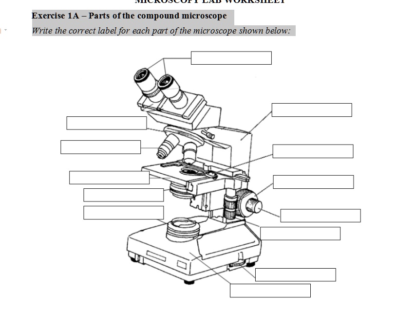
SOLVED: Exercise 1A _ Parts ofthe compound microscope Write ...
Fluorescence microscope - Wikipedia The majority of fluorescence microscopes, especially those used in the life sciences, are of the epifluorescence design shown in the diagram.Light of the excitation wavelength illuminates the specimen through the objective lens. The fluorescence emitted by the specimen is focused to the detector by the same objective that is used for the excitation which for greater resolution will …

Simple Microscope Definition, Magnification, Parts And Uses
depts.washington.edu › vurchinWelcome to Virtual Urchin - University of Washington Major update Apr 2021: All of the activities on the site are now mobile compatible !! Computers are still recommended, and tablets are preferable to phones: please read the Notes at the bottom of this page for details on the latest updates, mobile compatibility and general information about using this site.

General Biology | Carlson Stock Art | General biology ...
› ternary-phase-diagramTernary Phase Diagram - an overview | ScienceDirect Topics A point on the diagram represents a composition that is specified in terms of mole fraction or weight fraction. The point, (0.3, 0.4, 0.3) is at the center of the small triangle in the diagram and is located by following the red diagonal 60° line at red 0.3 and the horizontal line at blue 0.4 or any combination of two of the coordinates (A, B, C).

Microscope labeled diagram
Compound Microscope- Definition, Labeled Diagram, Principle, Parts, Uses The naked eye can now view the specimen at magnification 400 times greater and so microscopic details are revealed. Alternatively, the magnification of the compound microscope is given by: m = D/ fo * L/fe where, D = Least distance of distinct vision (25 cm) L = Length of the microscope tube fo = Focal length of the objective lens
Microscope, Microscope Parts, Labeled Diagram, and Functions
Simple Microscope - Diagram (Parts labelled), Principle, Formula and Uses Parts of a Simple Microscope A simple microscope consists of Optical parts Mechanical parts Labeled Diagram of simple microscope parts Optical parts The optical parts of a simple microscope include Lens Mirror Eyepiece Lens A simple microscope uses biconvex lens to magnify the image of a specimen under focus.

microscope drawing with label - Clip Art Library
Microscope, Microscope Parts, Labeled Diagram, and Functions 19.01.2022 · Revolving Nosepiece or Turret: Turret is the part of the microscope that holds two or multiple objective lenses and helps to rotate objective lenses and also helps to easily change power. Objective Lenses: Three are 3 or 4 objective lenses on a microscope. The objective lenses almost always consist of 4x, 10x, 40x and 100x powers. The most common eyepiece …

Label the microscope — Science Learning Hub
Label the microscope — Science Learning Hub 08.06.2018 · All microscopes share features in common. In this interactive, you can label the different parts of a microscope. Use this with the Microscope parts activity to help students identify and label the main parts of a microscope and then describe their functions.. Drag and drop the text labels onto the microscope diagram. If you want to redo an answer, click on the …

Microscope Parts worksheet
Electron microscope - Wikipedia An electron microscope is a microscope that uses a beam of accelerated electrons as a source of illumination. As the wavelength of an electron can be up to 100,000 times shorter than that of visible light photons, electron microscopes have a higher resolving power than light microscopes and can reveal the structure of smaller objects.. Electron microscopes use shaped magnetic …
Parts of the Microscope Labeling Activity!
Parts of a microscope with functions and labeled diagram 19.04.2022 · Figure: Diagram of parts of a microscope. There are three structural parts of the microscope i.e. head, base, and arm. Head – This is also known as the body. It carries the optical parts in the upper part of the microscope. Base – It acts as microscopes support. It also carries microscopic illuminators.

ABOUT MICROSCOPES | Scienceart
Neuron under Microscope with Labeled Diagram - AnatomyLearner But, first, let's try to identify the following features from a neuron with the help of a labelled diagram. Cell body or perikaryon of a neuron Nucleus, cytoplasm, the plasma membrane of a neuron Nissl bodies in the cell body of a neuron An initial segment of axon and axon hillock Dendrites and axons of a neuron Axolemma and myelin sheath

Parts of a microscope with functions and labeled diagram
Label The Parts Of A Microscope Worksheet Answers You can use the word bank below to fill in the blanks or cut. Label the parts of a microscope worksheet answers. Students label the parts of the microscope in this photo of a basic laboratory light microscope. Files include a link to editable doc so you can rewrite a. Power 10 x 4 40 Power 10 x 10 100 Power 10 x 40 400 What happens as the power ...

Cytology. Cytology. radiation used to illuminate the specimen ...
Welcome to Virtual Urchin - University of Washington microscope measurement. microscope compare. specimen compare. development & embryology. fertilization lab. embryogenesis to hatching. analyzing gene function. ecology & environment. our acidifying ocean. predator & prey. surfing to settlement. basic biology. urchin anatomy. about us. teacher resources. useful links . Select Language: Welcome to the new …
Scientific Tools Microscope Birth of the Microscope 1590
Electron Microscope-Definition, Principle, Types, Uses, Labeled Diagram Parts of an Electron Microscope The electron microscope is placed vertically and has the shape of a tall vacuum column. It consists of the following elements: 1. Electron gun A heated tungsten filament that produces electrons makes up the electron cannon. 2. Electromagnetic lenses The condenser lens directs the electron beam to the specimen.

This is a common compound microscope. Label its parts from A ...

Diagram of diatom microscope slide positioned with its label ...
Microscope Types (with labeled diagrams) and Functions
Microscope labeling, modern and classical types
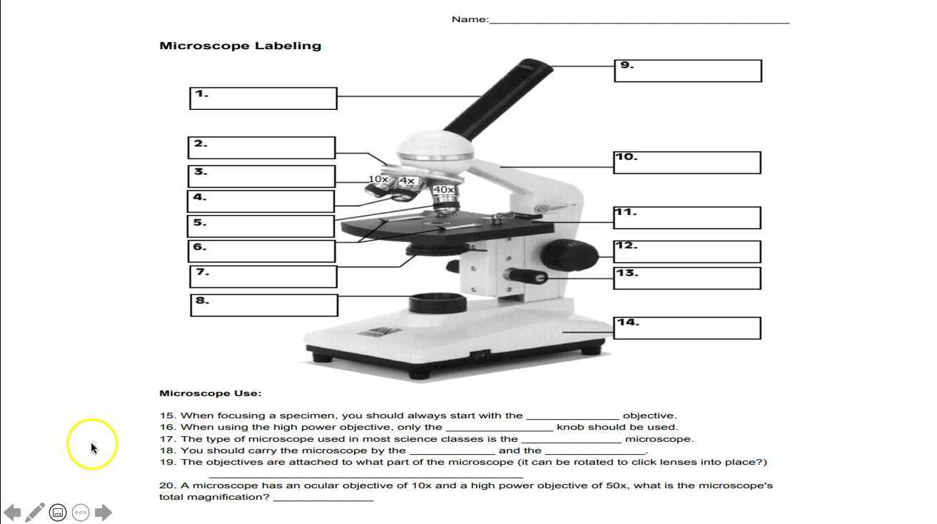
Microscope Labeling

Draw a labelled diagram of a compound microscope.
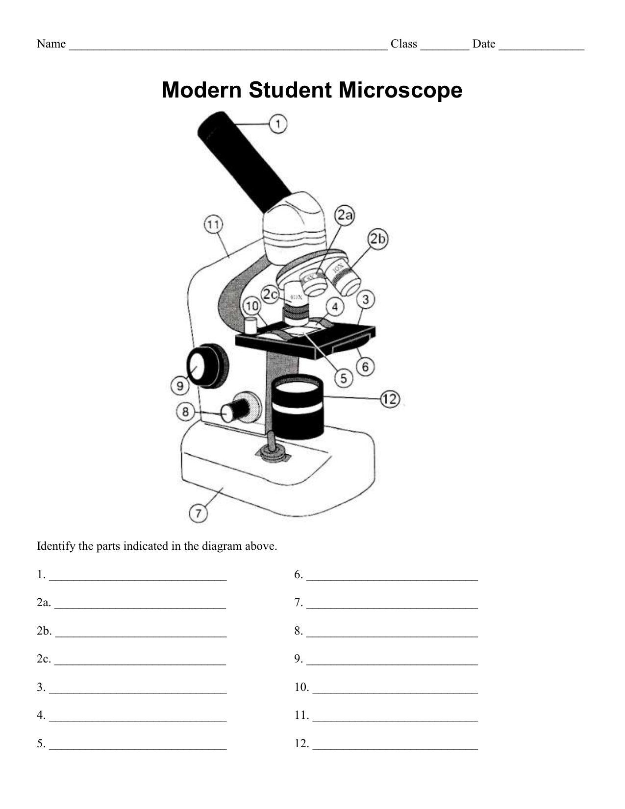
Name Date Sci STANDARD MICROSCOPE DIAGRAM Label only the ...
Instruments of Microscopy | Microbiology | | Course Hero
Mikroskop Binokular Biologi 40-5000X Set Portabel Percobaan Deteksi Ilmiah Perbesaran Tinggi Definisi Tinggi

Microscope Parts and Functions
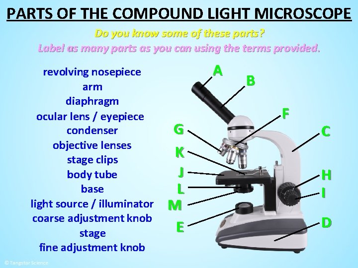
MICROSCOPE PARTS PARTS OF THE COMPOUND LIGHT MICROSCOPE

Microscope Labeling Activity - SMART Board Activity - Interactive Review
Microscope Diagram To Label - ClipArt Best - ClipArt Best ...
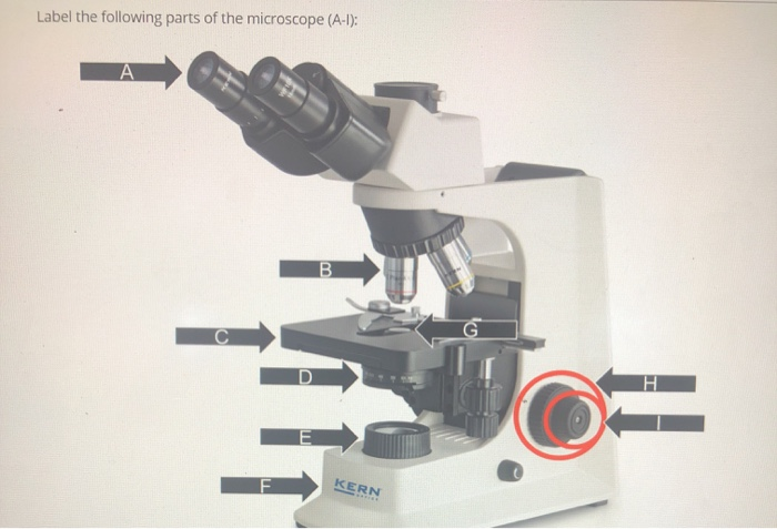
Solved Label the following parts of the microscope (A-1 ...

Lable the microscope worksheet

Microscope Labeling
Post a Comment for "44 label microscope diagram"