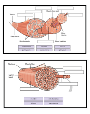
43 art-labeling activity: structure of muscle tissues
Answer correct art based question chapter 4 question - Course Hero ANSWER: Correctmultinucleate cells branched cells intercalated discs situated between cells striations tendons and ligaments attached to bones heart ducts of certain glands dense irregular connective tissue smooth muscle tissue skeletal muscle tissue cardiac muscle tissue art-labeling activity: figure 8.3 - markleyandvancamp Push your learning experience beyond the classroom with the Figure 83 Label Art Activity in the Introduction to Medical Terminology companion website. Figures 8-5 and 8-6 shows many of the muscles of the bodys trunk that you need to know as well as some of the muscles of the arms and legs you will learn about in the next lab.
Art-labeling Activity: Types of Connective Tissue Proper Art-labeling Activity: Types of Connective Tissue Proper 3.0 (2 reviews) + − Learn Test Match Created by leeny_montesino PLUS Terms in this set (6) Areolar Tissue ... Adipose Tissue ... Reticular Tissue ... Elastic Tissue ... Dense Irregular Connective Tissue ... Dense Regular Connective Tissue ... Sets found in the same folder

Art-labeling activity: structure of muscle tissues
exercise 13 review sheet art-labeling activity 3 Activities include organ dissections for rat cow and sheep specimens in addition to the full body dissections in the Cat and Pig versions of the lab manual giving students a hands-on lab experience. Fibularis brevis fibularis longus. Use the art-labeling activities to quiz yourself on key anatomical structures in this chapter. A & P Ch 6 Musclular System Student PPT - SlideShare Five Golden Rules of Skeletal Muscle Activity 1. With a few exceptions, all skeletal muscles cross at least one joint. 2. Typically, the bulk of a skeletal muscle lies proximal to the joint crossed. 3. All skeletal muscles have at least two attachments: the origin and the insertion. 4. Skeletal muscles can only pull; they never push. 5. art-labeling activity: components and divisions of the pelvis Instructors may assign this figure as an Art Labeling Activity using Mastering APTM Regional Anatomy The body is divided into two main regions the axial and appendicular regions. Curves and Regions of the Vertebral. Lesser pelvic cavity shape. 83 The Pelvic Girdle and Pelvis. School Greenfield Community College. Shape of the pelvic inlet.
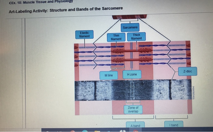
Art-labeling activity: structure of muscle tissues. Art Labeling Activity the Major Systemic Arteries - Blogger Identifying and Describing Skin Structures. The Major Systemic Arteris36 Terms. Arteries carry blood away from the heart in two distinct pathways. The major systemic arteries Part A Drag the appropriate labels to their respective targets. Brain cranium and meninges dural folds and sinuses Art-labeling Activity. art-labeling activity: the cell and its organelles The animal cell includes 17 organelles and the plant cell includes 20 organelles for students to label and color. Identify cell organelles on charts models and other laboratory material. The activity is a memorable experience for students to learn about the two cells by cutting coloring and creating their own plant or animal cell. [Solved] Art-labeling Activity: | Course Hero Answer to Art-labeling Activity: The Microscopic Structure of a Myofibril H band Zone of overlap M line A band Sarcomere Z line I band Submit Previous Answers ... Art-labeling Activity: ... A sarcomere is the area between two Z lines that can be regarded the fundamental structural and functional unit of muscle tissue. The Z-line establishes the ... Answered: Art-labeling Activity: Structure of the… | bartleby Science Anatomy and Physiology Q&A Library Art-labeling Activity: Structure of the testis Reset Help Seminiferous Ductus deferens - tubules Epididymis - Septa testis Efferent ductule - Mediastinum of testis Tunica vaginalis Skin of scrotum Scrotal cavity Tunica albuginea Straight tubule Cremaster Lobule Dartos muscle Rete testis.
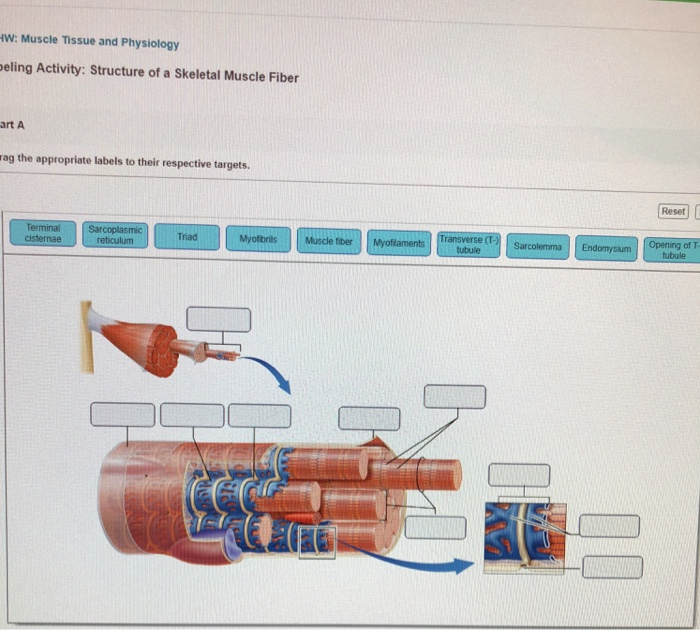
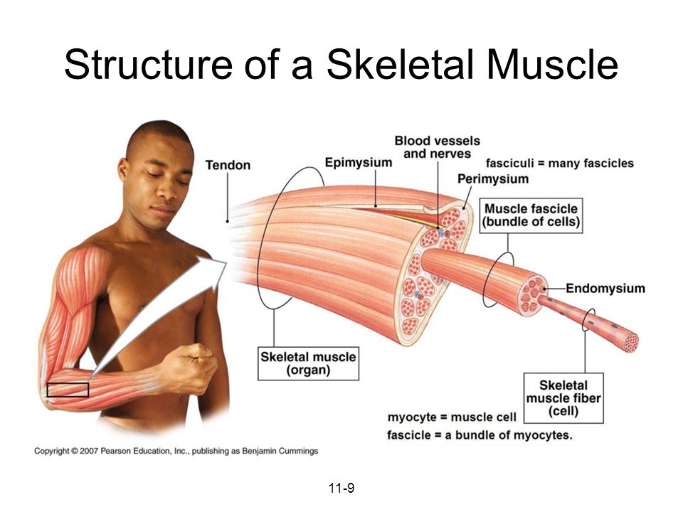
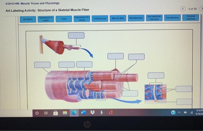
BIO 200 Chapter 9 - Muscle Tissue Physiology Flashcards | Quizlet When the sarcomere contracts and shortens__________. the A band stays the same. The storage and release of calcium ions is the key function of the: sarcoplasmic reticulum. A group of skeletal muscle fibers together with the surrounding perimysium form a (n): fascicle. Art-Ranking Activity: Stages of an action potential. A crossbridge forms when: [Solved] Art-Labeling Activity: | Course Hero Composed of actively dividing keratinocytes with spinous-like projections (prickle cells) This layer produces keratin and induces keratinization. Langerhans cells are also located in this layer. Stratum basale (also called the basal cell layer of the epidermis) Art- labeling Activity Flashcards | Quizlet Study with Quizlet and memorize flashcards containing terms like , , and more. Art-labeling Activity: The Structure of a Skeletal Muscle Fiber Start studying Art-labeling Activity: The Structure of a Skeletal Muscle Fiber. Learn vocabulary, terms, and more with flashcards, games, and other study tools.
chapter 9- Mastering A and P, Chapter 9-1 The muscular Tissue - Quizlet Art-labeling Activity: The structure of a skeletal muscle fiber PICTURE Chapter Test - Chapter 9 Question 3 Which thin-filament-associated structure is distinguished by its constituents of three globular subunits, one of which has a receptor that binds two calcium ions? a) G-actin b) nebulin c) tropomyosin d) troponin ... PDF Marieb HA8 chapter 4 - Pearson The word tissue derives from the Old French word meaning "to weave," reflecting the fact that the different tissues are woven together to form the "fabric" of the human body. The four basic types of tissue are epithelial tissue, connective tissue, muscle tissue, and nervous tissue. If a single, broad functional term were assigned to ... Answered: Art-labeling Activity: Structure of a… | bartleby Drag the labels to the appropriate location in the figure. Transcribed Image Text: Art-labeling Activity: Structure of a lymph node Medulla (B cells Cortex (B cells) and macrophages) Medullary sinus Medullary cord Paracortex (T cells) Efferent vessel Capsule Hilum Subcapsular Afferent vessel space Lymph node artery and vein Trabeculae • Previous. art labeling activity the heart wall - jacobvaneycksheetmusic Students must label the parts of the heart including. Internal Anatomy of the Heart heart wall and the pericardium Art-labeling. Layers Of The Uterine Wall. Use the art-labeling activities to quiz yourself on key anatomical structures in this chapter. Drag the labels tothe appropriate location in the figure. School Miami Dade College Miami.
Muscle Tissue: An Introduction. Muscles make up close to half ...
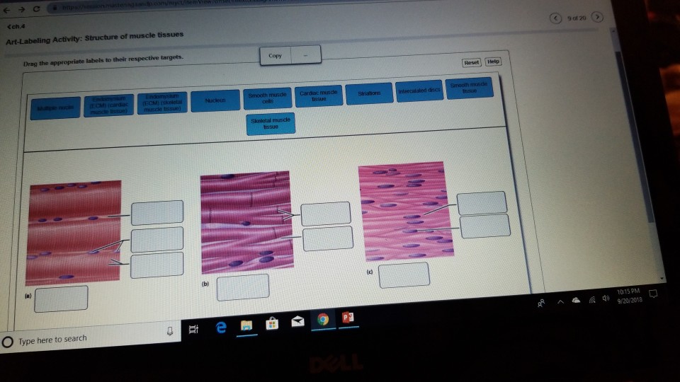
Tissues Lab p5-8.pdf - Reset Loose CT Hyaline cartilage... Correct The structure labeled D is a collagen fiber. Collagen fibers are thick, and appear pink due to staining. Art-Labeling Activity: Structure of muscle tissues Part A Drag the appropriate labels to their respective targets.
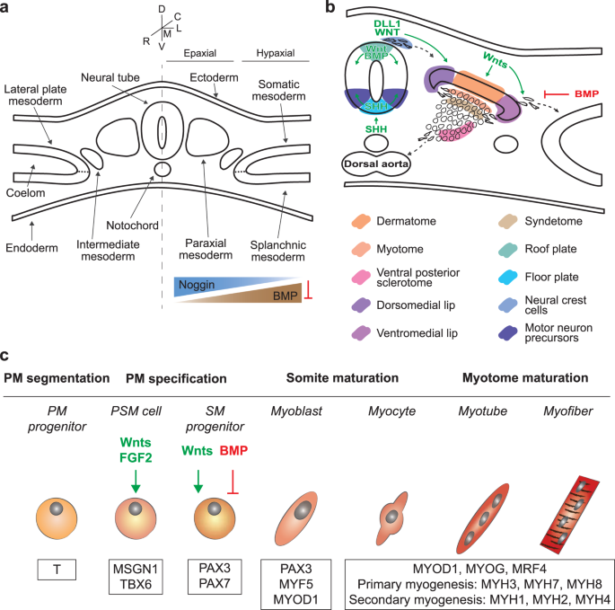
Skeletal muscle differentiation of human iPSCs meets ...
Answered: Art-labeling Activity: Structural… | bartleby Q: Label different areas of an individual muscle unit known as a sarcomere below: Actin A Band Mine… A: The smallest functional unit of muscle tissue is called sarcomere. It consist of actin and myosin…
Skeletal muscle tissue: Histology | Kenhub
Muscles Labeling - The Biology Corner Muscles Labeling. Shannan Muskopf November 11, 2020. This activity is aligned to my anatomy and physiology curriculum where students study the structure and function of muscle tissues. This has been a challenging topic to cover remotely because I can't use traditional models. Typically, I would use straws and rubber bands to model fascicles ...

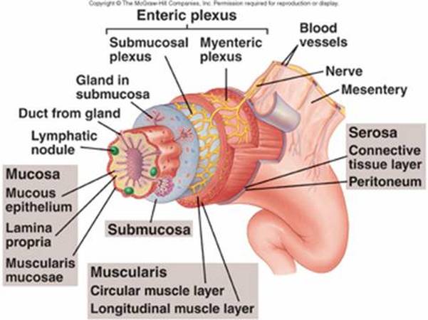
Art-labeling Activity: Diagrammatic sectional view along the ...
Part a muscle tissue has the ability to contract when Correct Art-labeling Activity: The Organization of Skeletal Muscles (1 of 2) Drag the labels onto the diagram to identify structural features associated with skeletal muscle. Part A Drag the labels onto the diagram to identify structural features associated with skeletal muscle.

Skeletal System - ScienceDirect
Solved Secure https:/ C Lab: Histology Art-labeling | Chegg.com Question. : Secure https:/ C Lab: Histology Art-labeling Activity: Structure of muscle tissues 102091378 Part A Drag the appropriate labels to their respective targets tissue Smooth muSECM) (cardiac tissue (ECM) (skeletal Nucleus issue Type here to search.
A Functional Approach to Human Anatomy Available in a ...
Answered: Art-labeling Activity: Structural… | bartleby Transcribed Image Text:

Single center, open label dose escalating trial evaluating ...
art-labeling activity: components and divisions of the pelvis Instructors may assign this figure as an Art Labeling Activity using Mastering APTM Regional Anatomy The body is divided into two main regions the axial and appendicular regions. Curves and Regions of the Vertebral. Lesser pelvic cavity shape. 83 The Pelvic Girdle and Pelvis. School Greenfield Community College. Shape of the pelvic inlet.

Taste receptor - Wikipedia
A & P Ch 6 Musclular System Student PPT - SlideShare Five Golden Rules of Skeletal Muscle Activity 1. With a few exceptions, all skeletal muscles cross at least one joint. 2. Typically, the bulk of a skeletal muscle lies proximal to the joint crossed. 3. All skeletal muscles have at least two attachments: the origin and the insertion. 4. Skeletal muscles can only pull; they never push. 5.
Solved Art-labeling activity: structure of skeletal muscle ...
exercise 13 review sheet art-labeling activity 3 Activities include organ dissections for rat cow and sheep specimens in addition to the full body dissections in the Cat and Pig versions of the lab manual giving students a hands-on lab experience. Fibularis brevis fibularis longus. Use the art-labeling activities to quiz yourself on key anatomical structures in this chapter.
A & P Ch 6 Musclular System Student PPT

Normal Muscle Fiber Stock Illustrations – 52 Normal Muscle ...
Solved] Art-Labeling Activity: Main effects of sympathetic ...
In vitro drug testing based on contractile activity of C2C12 ...
Solved
Muscles and Muscle Tissue
Protein synthesis rates of muscle, tendon, ligament ...
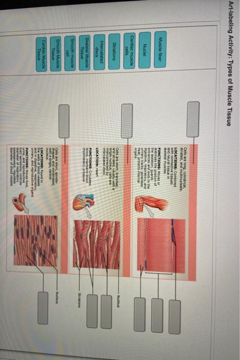
Solved Art-labeling Activity: Types of Muscle Tissue | Chegg.com
Solved (-ラC cch.4 Art-Labeling Activity: Structure of muscle ...
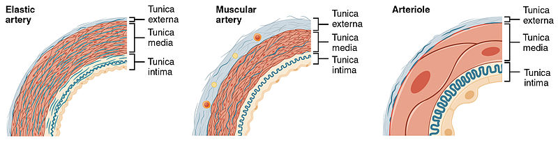
What is the Difference Between Elastic and Muscular Arteries ...

Art-labeling Activity: Types of Connective Tissue Proper ...
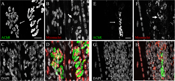
Exclusive vital labeling of myonuclei for studying myonuclear ...
Anatomy and Physiology Lab I” on OpenALG
Muscles Labeling

Art-labeling Activity: The Structure of a Skeletal Muscle ...
OVERVIEW OF MUSCLE TISSUE

BIO 200 Chapter 9 - Muscle Tissue Physiology Flashcards | Quizlet
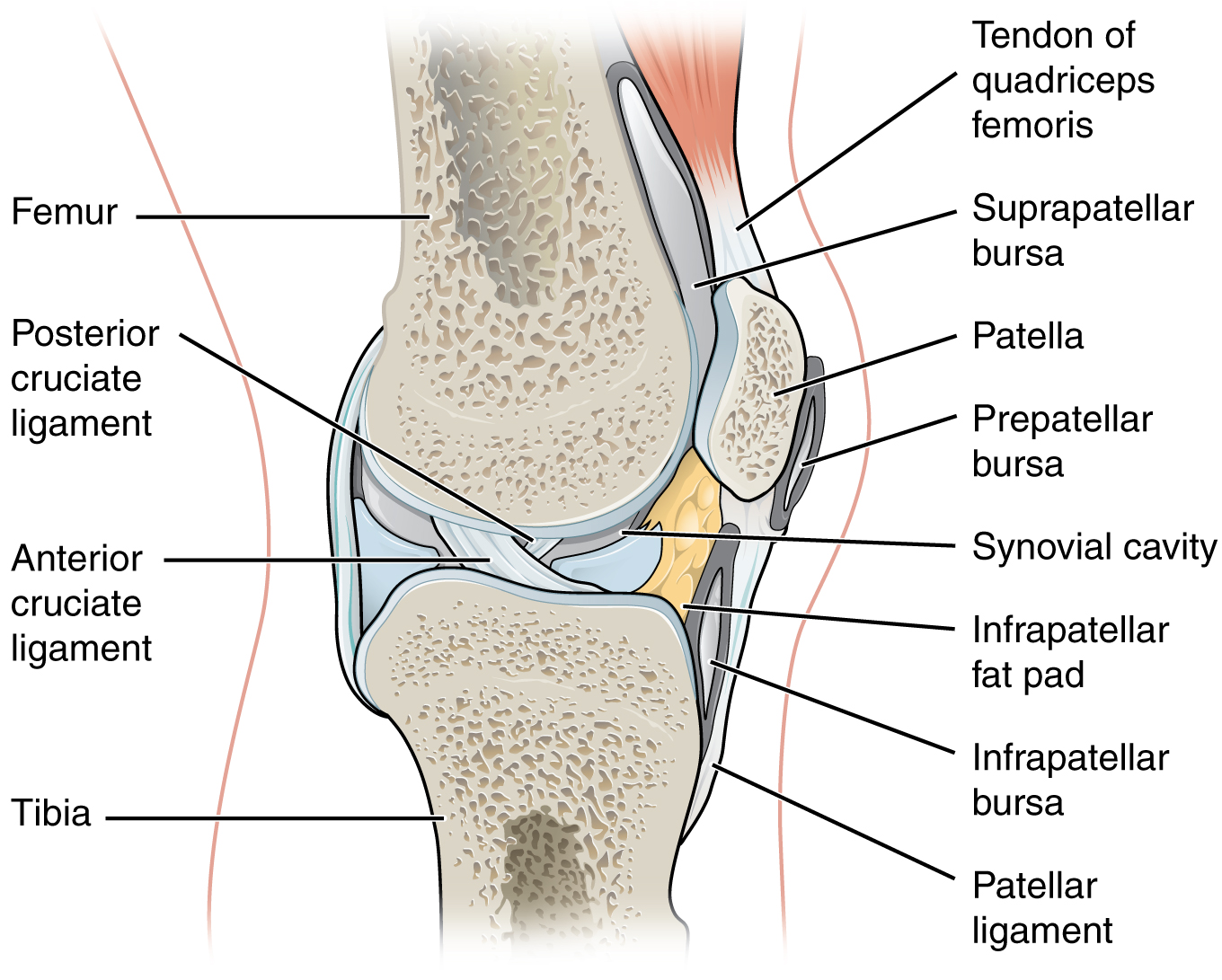
Synovial Joints – Anatomy & Physiology
Muscles. Skeletal Muscle Introduction Movement is a ...
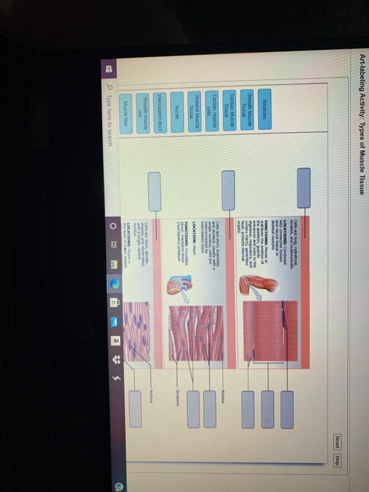
Solved Art-labeling Activity: Types of Muscle Tissue Reset ...
HistologyCh4 Student handoutF15.pptx
11 Introduction to the Nervous System and Nervous Tissue
Nervous Tissue

BIO 200 Chapter 9 - Muscle Tissue Physiology Flashcards | Quizlet

What is the function of osteocalcin? - ScienceDirect
Ch. 7
What does luminal mean in anatomy? - Quora
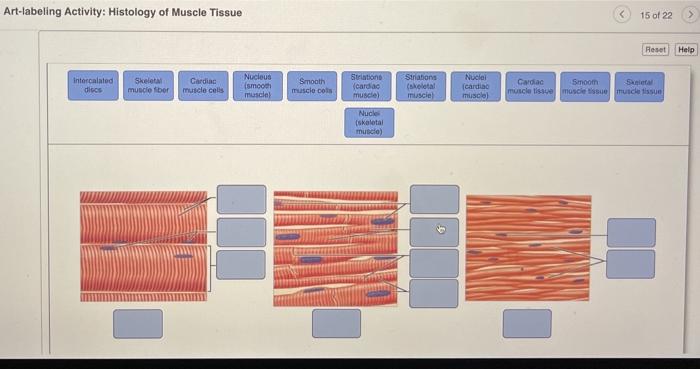
Solved Art-labeling Activity: Histology of Muscle Tissue 15 ...
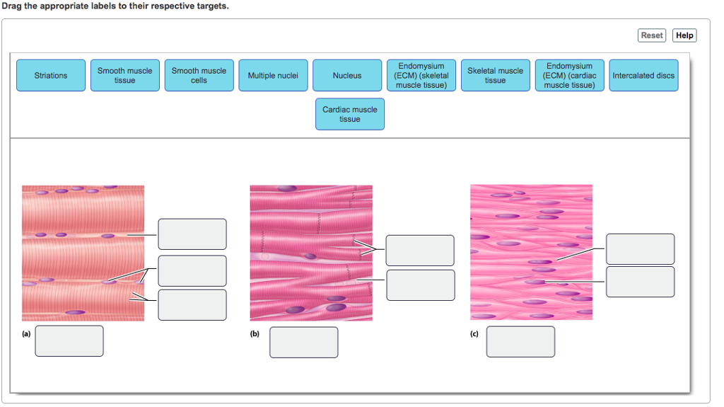
Solved Drag the appropriate labels to their respective ...
Blog Archives - The human body
Major structures of the sole of the foot, inferior view ...
Solved

Anatomy of the Breast, Axilla, and Chest Wall | Oncohema Key
Muscles and Muscle Tissue
Post a Comment for "43 art-labeling activity: structure of muscle tissues"