39 phospholipid labeled
Label the Phospholipid Bilayer Diagram | Quizlet phospholipid composed of a hydrophobic tail and a hydrophilic head hydrophilic heads Negative charge so they attract to water hydrophobic tails Fatty acids are nonpolar and hydrophobic cholesterol maintain fluidity of the membrane and prevent non polar fatty acid tails from sticking together even in cold temperatures peripheral protein Home Page: Thrombosis Research Sep 07, 2022 · We use cookies to help provide and enhance our service and tailor content. To update your cookie settings, please visit the Cookie Preference Center for this site.
Phospholipid Structure Labeling Diagram | Quizlet Phospholipid Structure Labeling + − Learn Test Match Created by njabara Terms in this set (6) Phosphate ... Glycerol ... Saturated Fatty Acid ... Unsaturated Fatty Acid ... Hydrophobic Tails ... Hydrophilic Head ... Sets found in the same folder types of solutions 7 terms Mimi853 (Ch. 3) Plasma Membrane (cell membrane) 19 terms

Phospholipid labeled
Quanta BioDesign Quanta BioDesign's building blocks and protected linkers enable SuperHydrophilic™ constructs and peptide modifications via our world-famous dPEG ® products. Our simple-to-use products transform hydrophobic peptides into hydrophilic products. Phospholipid - Wikipedia Phospholipids, [1] are a class of lipids whose molecule has a hydrophilic "head" containing a phosphate group and two hydrophobic "tails" derived from fatty acids, joined by an alcohol residue (usually a glycerol molecule). Marine phospholipids typically have omega-3 fatty acids EPA and DHA integrated as part of the phospholipid molecule. [2] Phospholipid structure (video) | Khan Academy There's phosphatidylserine, phosphatidylcholine, phosphatidtylethanolomine, phosphatidylinositol, and diphosphatidylgylcerol, also known as cardiolipin. And you'll notice that in this last one, there are actually two phosphatidyl p groups that actually bond to a middle gylcerol.
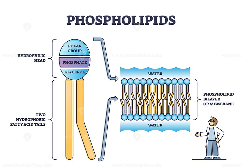
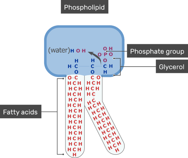
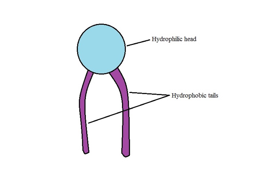
Phospholipid labeled. Phospholipid: Definition, Structure, Function, Examples - Science Terms Phospholipid Definition A phospholipid is an amphiphilic molecule consisting of a polar head region, a unit of glycerol, and two or more non-polar fatty acid tails, typically found in a cell membrane. A bilayer of phospholipid molecules forms a plasma membrane. Phospholipid: Definition, Structure, Function | Biology Dictionary Phospholipid Definition A phospholipid is a type of lipid molecule that is the main component of the cell membrane. Lipids are molecules that include fats, waxes, and some vitamins, among others. Each phospholipid is made up of two fatty acids, a phosphate group, and a glycerol molecule. Plant Cell - The Definitive Guide | Biology Dictionary 15/01/2021 · Labeled diagram of a chloroplast Vacuoles. Plant cells are unique in that they have a large central vacuole. A vacuole is a small sphere of plasma membrane within the cell that can contain fluid, ions, and other molecules. Vacuoles are essentially just large vesicles. They can be found in the cells of many different organisms. However, plant cells characteristically have a … Directionality (molecular biology) - Wikipedia Directionality, in molecular biology and biochemistry, is the end-to-end chemical orientation of a single strand of nucleic acid.In a single strand of DNA or RNA, the chemical convention of naming carbon atoms in the nucleotide pentose-sugar-ring means that there will be a 5′ end (usually pronounced "five-prime end"), which frequently contains a phosphate group attached to the 5′ carbon of ...
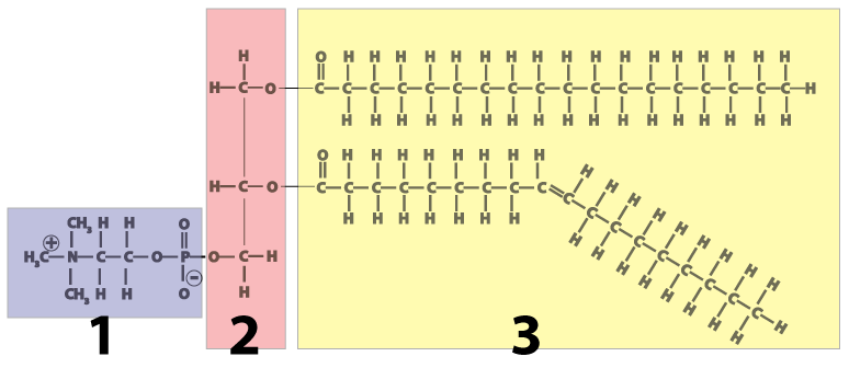
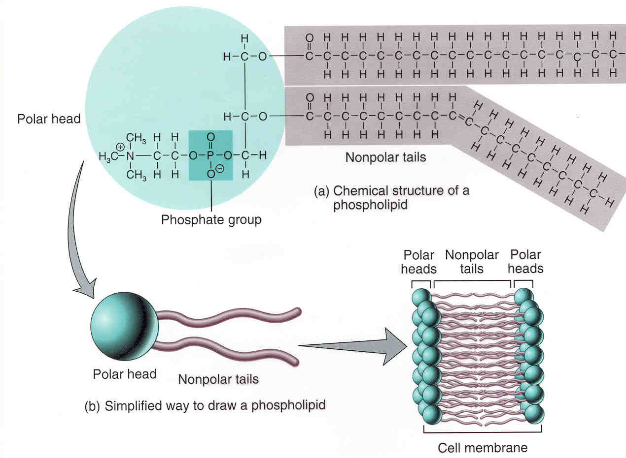
Fatty Acid Analogs and Phospholipids—Section 13.2 The spectral properties of BODIPY FL dye-labeled phospholipids are summarized in Spectral properties of some lipid probes—Table 13.2. Unlike the nitrobenzoxadiazole (NBD) fluorophore, the BODIPY FL and BODIPY 500/510 fluorophores are intrinsically lipophilic and readily localize in the membrane's interior. Phospholipids | Introduction to Chemistry | | Course Hero Phospholipid Molecule A phospholipid is a molecule with two fatty acids and a modified phosphate group attached to a glycerol backbone. The phosphate may be modified by the addition of charged or polar chemical groups. Two chemical groups that may modify the phosphate, choline and serine, are shown here. F-ATPase - Wikipedia F-ATPase, also known as F-Type ATPase, is an ATPase/synthase found in bacterial plasma membranes, in mitochondrial inner membranes (in oxidative phosphorylation, where it is known as Complex V), and in chloroplast thylakoid membranes.It uses a proton gradient to drive ATP synthesis by allowing the passive flux of protons across the membrane down their … Phospholipids - Structure, Types, Properties and Function - VEDANTU A phospholipid is a molecule containing a glycerol backbone and two fatty acids linked, as well as a modified phosphate group. The addition of charged or polar chemical groups to the phosphate can change its properties. Choline and serine, two chemical groups that can alter phosphate.
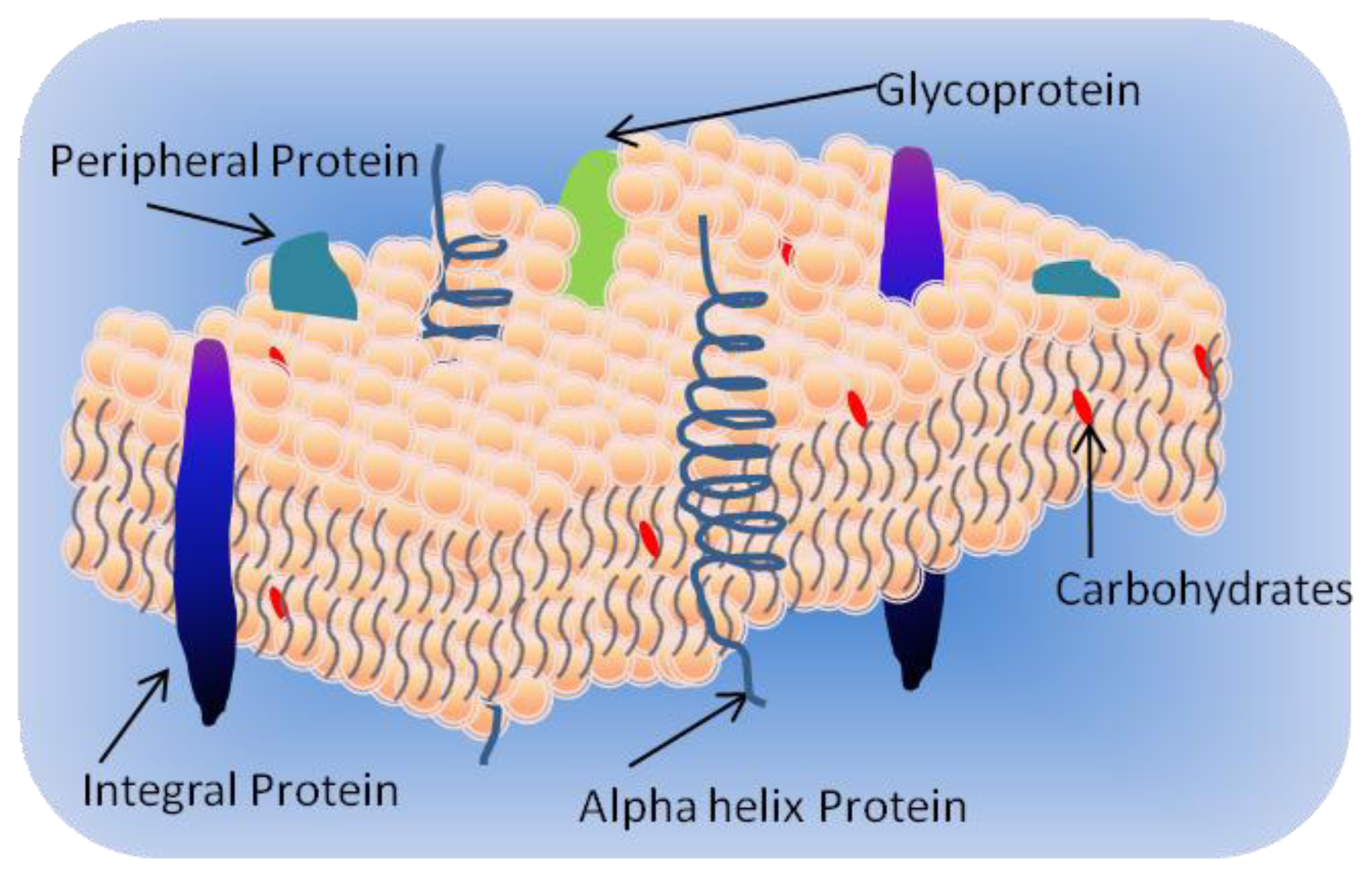
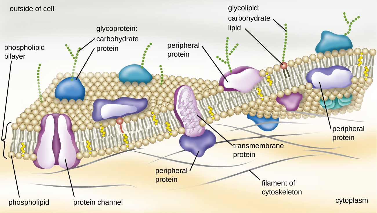
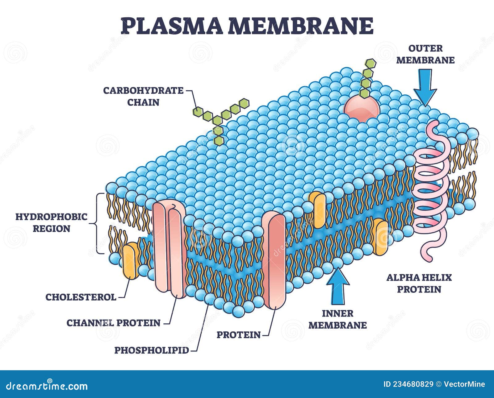
Labeling phospholipid membranes with lipid mimetic luminescent metal ... Two lipid-mimetic luminescent labels have been introduced and fully characterized. • The labels are highly photostable with large Stokes shift and up to 888 ns lifetime. • The labels are readily incorporated into neat DMPC as well as mixed lipid membranes. • The labels do not alter the physicochemical properties of biomimetic membranes. Abstract Sandwich (Davson–Danielli) model of cell membrane - Microbe … 06/04/2022 · Danielli and Davson proposed a model whereby two layers of protein flanked a central phospholipid bilayer. The model was also described as a ‘lipo-protein sandwich’, as the lipid layer was sandwiched between two protein layers. The Davson–Danielli model predominated until Singer and Nicolson advanced the fluid mosaic model in 1972. The Lipid Bilayer - Molecular Biology of the Cell - NCBI Bookshelf The motion and orientation of a spin-labeled lipid in a bilayer can be deduced from the ESR spectrum. Such studies show that phospholipid molecules in synthetic bilayers very rarely migrate from the monolayer (also called a leaflet) on one side to that on the other. This process, known as “flip-flop,” occurs less than once a month for any ... Cells - University of Utah In multicellular organisms, cells work together in teams. Multiple cell types, each specialized for a certain function, team up to form tissues.
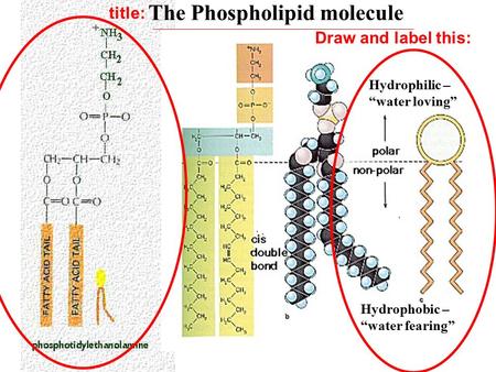
Describing the Structure and Nature of a Phospholipid
Phospholipids Teaching Resources | Teachers Pay Teachers Simple, effective label resource!Included for this PHOSPHOLIPID BILAYER activity:Label sheetLabel sheet with keywordsLabel sheet answersThis activity focusses on keywords, with a bold image to label. The document is completely editable in PowerPoint but also comes with a PDF copy for preparation free printing. Every page included in the ...

Chemical Structure of Lipids — Overview & Types - Expii
which part of a phospholipid is hydrophobic - solsarin Phospholipid Molecule A phospholipid is a molecule with two fatty acids and a modified phosphate group attached to a glycerol backbone. The phosphate may be modified by the addition of charged or polar chemical groups. Two chemical groups that may modify the phosphate, choline and serine, are shown here.

Phospholipid - Definition and Examples - Biology Online ...
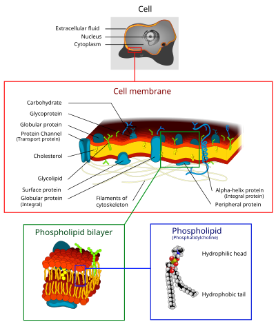
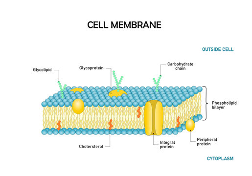
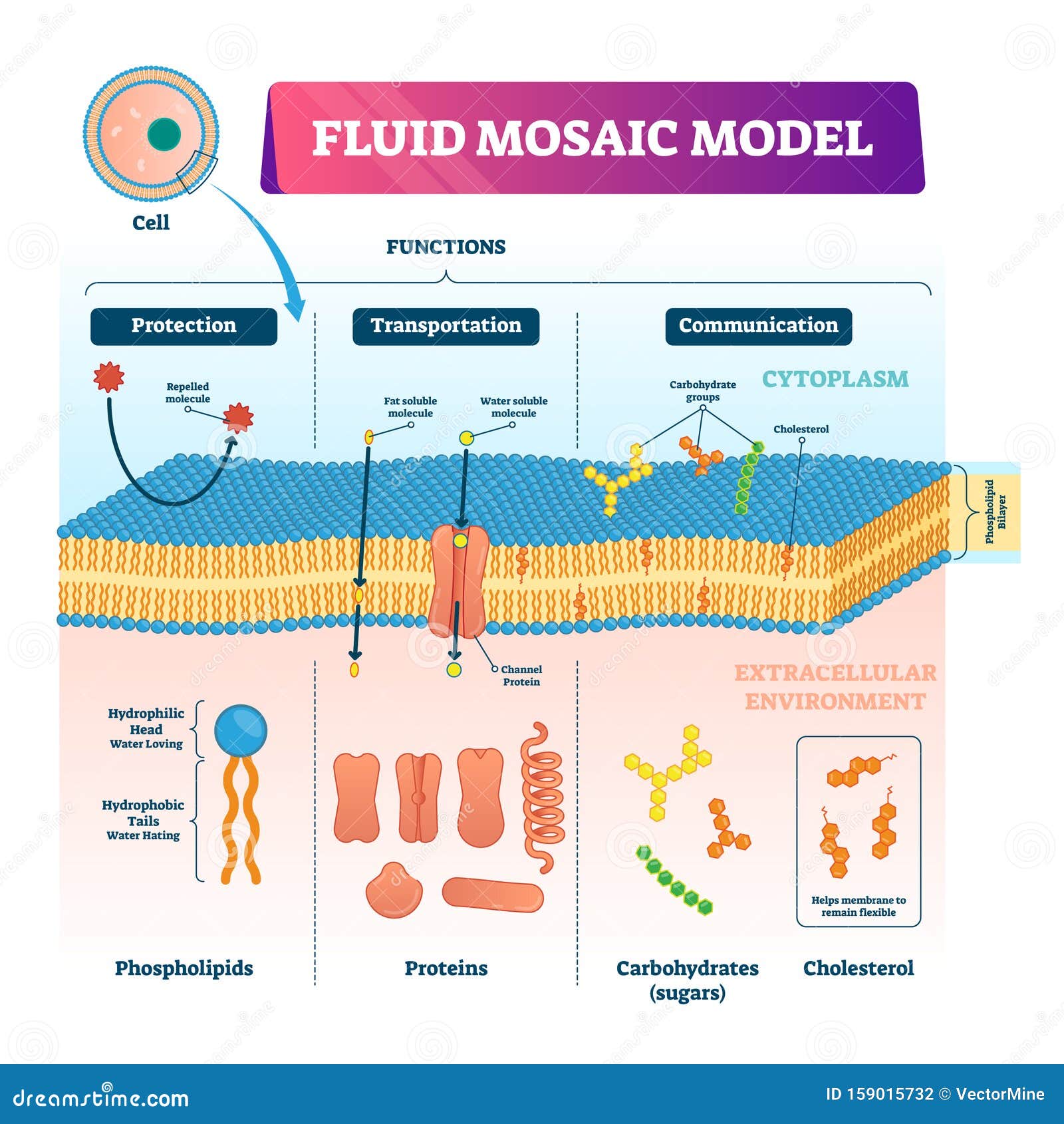
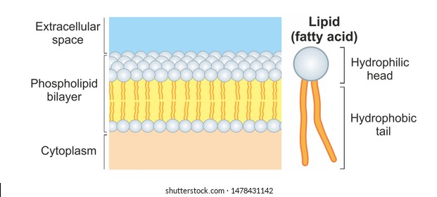
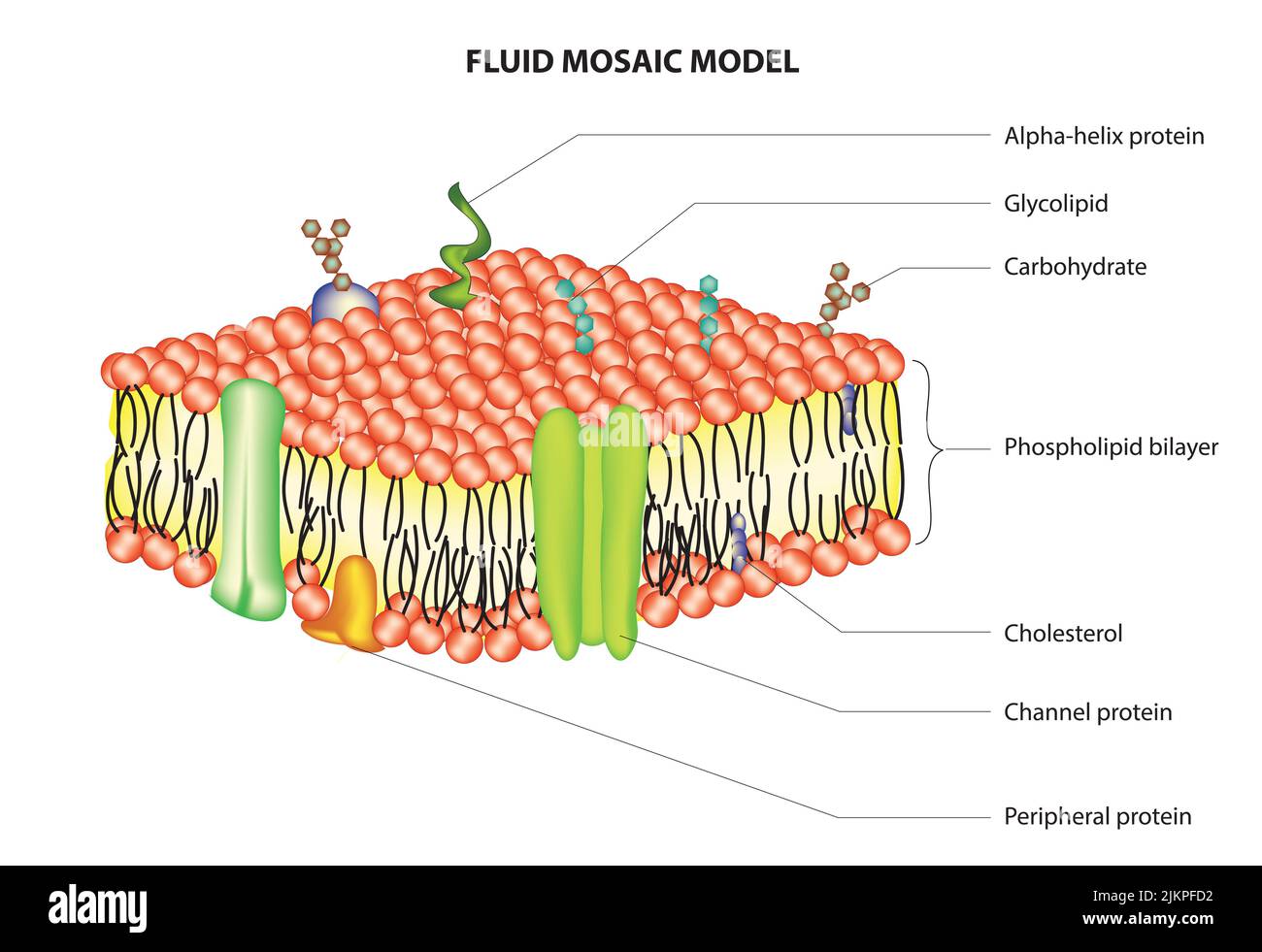
Phospholipid Bilayer | Lipid Bilayer | Structures & Functions Phospholipid Bilayer: All cells are surrounded by the cell membranes, and this characteristic best portrayed by the Fluid Mosaic Model. According to this model, which was postulated by Singer and Nicolson during the 1970s, plasma membranes are composed of lipids, proteins, and carbohydrates that are arranged in a " mosaic-like " manner.
Phospholipid or phosphatides lipids head and tail structure outline diagram
Metabolic labeling and direct imaging of choline phospholipids ... - PNAS Phospholipid molecules bearing a terminal alkyne group can be detected by "click" chemistry using a fluorescent azide. ( B) Propargyl-Cho incorporation into NIH 3T3 cells. Cells labeled overnight with varying concentrations of propargyl-Cho were fixed and stained with Alexa568-azide.
IJMS | Free Full-Text | Engineering Lipid Bilayer Membranes ...
Lipophilic Tracers—Dil, DiO, DiD, DiA, and DiR - Thermo Fisher … physiological properties.1,2 DiI-labeled moto neurons reportedly have remained viable for up to four weeks in culture and up to one year in vivo.5 The dyes uniformly label neurons via lateral diffusion in the plasma membrane at a rate of about 0.2–0.6 mm per day in fixed speci-
Phospholipid Vector Art Stock Images | Depositphotos
Phospholipid Bilayer | Introduction, Structure and Functions - iBiologia Phospholipid Definition. Phospholipid Bilayer is basically a special form of lipid molecule which is mainly the major constituent of the Cell Membrane. Fats, Waxes, and Vitamins are the molecules that are Lipids in nature and composed of Lipids. While Phospholipid is comprised of two molecules of Fatty acids, Phosphate Group, and a Glycerol ...
Membranes Interactive Tutorial 1: The Phospholipid Bilayer ...
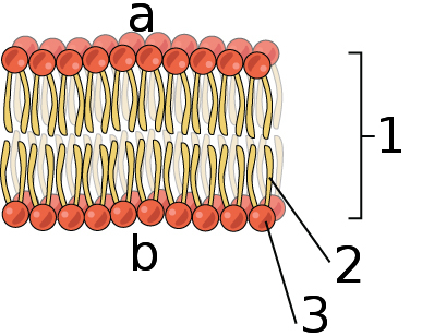
Phospholipid Bi-Layer Diagram - SmartDraw Phospholipid Bi-Layer Diagram Create Biology Diagram examples like this template called Phospholipid Bi-Layer Diagram that you can easily edit and customize in minutes. 7/20 EXAMPLES EDIT THIS EXAMPLE Text in this Example: Na- Phospholipid Bi-layer (Potasium Ion Channel example) Cytoplasm Sodium Ion Channel Potassium Ion K+ Phospholipid CH CH2 CH3

3.5: Lipid Molecules - Phospholipids - Biology LibreTexts
Solved Plasma Membrane Assignment Draw and label a section - Chegg Plasma Membrane Assignment. Draw and label a section of the plasma membrane of a human cell. Your drawing should include: A phospholipid bilayer: Each phospholipid should include at least one unsaturated, fatty acid tail. Your plasma membrane should be at least 15-20 phospholipids in length. Label the outside surface and the inside surface of ...
Cell Membrane Lipid Bilayer | GetBodySmart
Metabolic labeling and direct imaging of choline phospholipids ... - PubMed The resulting propargyl-labeled phospholipid molecules can be visualized with high sensitivity and spatial resolution in cells via a Cu (I)-catalyzed cycloaddition reaction between the terminal alkyne group of propargyl-Cho and a labeled azide.
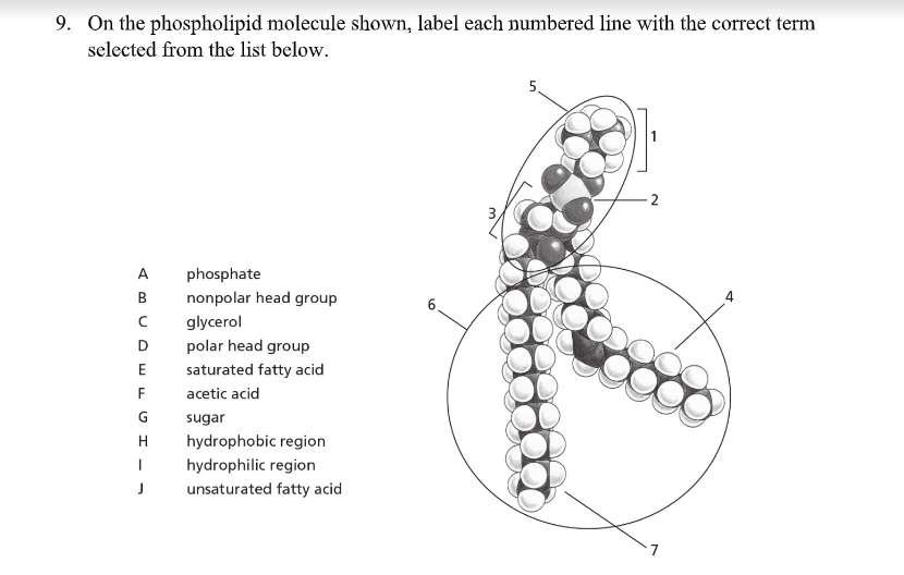
Solved 9. On the phospholipid molecule shown, label each ...
Lipids - Michigan State University If a phospholipid is smeared over a small hole in a thin piece of plastic immersed in water, a stable planar bilayer of phospholipid molecules is created at the hole. As shown in the following diagram, the polar head groups on the faces of the bilayer contact water, and the hydrophobic alkyl chains form a nonpolar interior. The phospholipid molecules can move about in their half …
Cell membrane - Wikipedia
In Vivo Tracking of Multiple Tumor Exosomes Labeled by Phospholipid ... In Vivo Tracking of Multiple Tumor Exosomes Labeled by Phospholipid-Based Bioorthogonal Conjugation Anal Chem. 2018 Oct 2 ;90(19 ... This phospholipid-based labeling strategy opens a new window to directly visualize and monitor exosome trafficking in living systems and holds great promise for exploring exosome-involved biological events such as ...
Phospholipid Images – Browse 1,375 Stock Photos, Vectors, and ...
Flip-flop of fluorescently labeled phospholipids in proteoliposomes ... Proteoliposomes prepared from a Triton X-100 extract of yeast microsomal membranes were also capable of flipping NBD-labeled phospholipid analogues rapidly in an ATP-independent fashion. Flippase activity was sensitive to the protein modification reagents N-ethylmaleimide and diethylpyrocarbonate.

Cell Membrane: Functions, Role & Structure - Video & Lesson ...
Phospholipid structure (video) | Khan Academy There's phosphatidylserine, phosphatidylcholine, phosphatidtylethanolomine, phosphatidylinositol, and diphosphatidylgylcerol, also known as cardiolipin. And you'll notice that in this last one, there are actually two phosphatidyl p groups that actually bond to a middle gylcerol.

Phospholipid Structure & Function | What is a Phospholipid ...
Phospholipid - Wikipedia Phospholipids, [1] are a class of lipids whose molecule has a hydrophilic "head" containing a phosphate group and two hydrophobic "tails" derived from fatty acids, joined by an alcohol residue (usually a glycerol molecule). Marine phospholipids typically have omega-3 fatty acids EPA and DHA integrated as part of the phospholipid molecule. [2]

Lipids | Microbiology
Quanta BioDesign Quanta BioDesign's building blocks and protected linkers enable SuperHydrophilic™ constructs and peptide modifications via our world-famous dPEG ® products. Our simple-to-use products transform hydrophobic peptides into hydrophilic products.
Phospholipid Bilayer | Introduction, Structure and Functions

Phospholipid Diagram Diagram | Quizlet

Phospholipid structures. The diagram shows the structures of ...

Lipids | Biology for Non-Majors I
5.4: Plasma Membrane - Biology LibreTexts

Binding of NBD-labeled phospholipid and sphingolipid probes ...

File:0301 Phospholipid Structure labeled.jpg - Wikimedia Commons
Fluid Mosaic Model Vector Illustration. Cell Membrane ...

Phospholipid Structure Labeling Diagram | Quizlet

Internalization of NBD-labeled phospholipid analogues in ...

Phospholipid bilayer composed of hydrophobic non-polar tails ...
Cell Membrane - Definition, Function/Structure, Animal/Plant cell

Media Portfolio
Phospholipid Bilayer. The Cell Membrane a phospholipid ...
1,649 Phospholipids Images, Stock Photos & Vectors | Shutterstock

Learn About Structure Of Phospholipid | Chegg.com
Solved] Draw and label diagram of a cell membrane, showing ...

Fluid mosaic model: cell membranes article (article) | Khan ...
Cell Membrane or Cytoplasmic Membrane Microscopic Structure ...

Cell Membrane Function and Structure
Vector Bilayer Stock Illustrations – 103 Vector Bilayer Stock ...
Phospholipid hi-res stock photography and images - Alamy
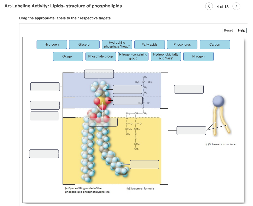
Solved Art-Labeling Activity: Lipids- structure of | Chegg.com
File:0302 Phospholipid Bilayer labeled.jpg - Wikimedia Commons
Post a Comment for "39 phospholipid labeled"