43 label the parts of a compound microscope
PDF Biological Diagram Of Simple Microscope With Label The Parts of a Microscope Labeled Printable TeacherVision bespoke.cityam.com 5 / 12. Biological Diagram Of Simple Microscope With Label June 22nd, 2018 - This diagram labels and explains the function of each part of a microscope Use this printable as a handout or transparency to help prepare students for working with ... Biological Diagram Of ... Label Parts Of A Compound Microscope Teaching Resources | TPT This is a set of 3 tiered readings. Students will read a passage about the how to use a compound light microscope. Students will use textual evidence to answer questions and label the different parts of the microscope. It also allows students to gain prior knowledge about the compound microscope. Version A provides the most support for students.
Compound Microscope: Definition, Diagram, Parts, Uses, Working Principle The compound microscope is mainly used for studying the structural details of cell, tissue, or sections of organs. The parts of a compound microscope can be classified into two: Non-optical parts Optical parts Non-optical parts Base The base is also known as the foot which is either U or horseshoe-shaped.

Label the parts of a compound microscope
Microscopy: Intro to microscopes & how they work (article) - Khan Academy Magnification is a measure of how much larger a microscope (or set of lenses within a microscope) causes an object to appear. For instance, the light microscopes typically used in high schools and colleges magnify up to about 400 times actual size. So, something that was 1 mm wide in real life would be 400 mm wide in the microscope image. 16 Parts of a Compound Microscope: Diagrams and Video The 16 core parts of a compound microscope are: Head (Body) Arm Base Eyepiece Eyepiece tube Objective lenses Revolving Nosepiece (Turret) Rack stop Coarse adjustment knobs Fine adjustment knobs Stage Stage clips Aperture Illuminator Condenser Diaphragm Video: Parts of a compound Microscope with Diagram Explained Compound Microscope Labeled Diagram | Quizlet Compound Microscope Labeled + − Flashcards Learn Test Match Created by meganplocher734 Terms in this set (14) Eyepiece/Ocular lens Contains the ocular lens Body tube A hollow cylinder that holds the eyepiece. Arm Part that supports the microscope. Stage Supports the slide or specimen Coarse adjustment Knob
Label the parts of a compound microscope. Microscope Parts Labeling Worksheets Parts of a microscope labeling the parts of a compound microscope id: Web if so, this parts of a light microscope activity is a great way of showing your students all of the different parts that go into making one!by breaking up the microscope into different. Source: studylib.net. Microscope | Biology I Laboratory Manual / Lab Report #2 - Learning How ... Another microscope that him will use in lab is a static otherwise a dissecting microscope. This sort of microscope user visible light view thicker, larger specimens, how as an insect, inside 3D. Since i are display larger samples, the magnification distance of this dissecting microscope is lower for the compound light microscope. Parts of a Microscope - SmartSchool Systems / Microscope Worksheet Labeled Microscope Parts Worksheets Slide - JPG Word Document PDF Designated Parts of adenine Microscope Image Labeled Parts of a Microscope Word Document Labeled Parts of a Microscope PDF Unlabeled Microscope Parts Worksheets Image - JPG Word Document PDF Unlabeled Parts of a Microscope Image Unlabeled Parts of a Microscope Word Download Unlabeled Body of … Continue reading Spare of a ... Compound Microscope Parts The three basic, structural components of a compound microscope are the head, base and arm. Head/Body houses the optical parts in the upper part of the microscope Base of the microscope supports the microscope and houses the illuminator Arm connects to the base and supports the microscope head. It is also used to carry the microscope.
Label the microscope — Science Learning Hub Label the microscope Interactive Add to collection Use this interactive to identify and label the main parts of a microscope. Drag and drop the text labels onto the microscope diagram. diaphragm or iris base eye piece lens fine focus adjustment light source stage coarse focus adjustment high-power objective Download Exercise Parts of a Microscope with Their Functions • Microbe Online The common light microscope used in the laboratory is called a compound microscope. It is because it contains two types of lenses; ocular and objective. The ocular lens is the lens close to the eye, and the objective lens is the lens close to the object. These lenses work together to magnify the image of an object. Parts of Compound Microscope Compound Microscope: Parts of Compound Microscope - BYJU'S The parts of the compound microscope can be categorized into: Mechanical parts Optical parts (A) Mechanical Parts of a Compound Microscope 1. Foot or base It is a U-shaped structure and supports the entire weight of the compound microscope. 2. Pillar It is a vertical projection. This stands by resting on the base and supports the stage. 3. Arm Label Parts of a Compound Light Microscope Flashcards Study with Quizlet and memorize flashcards containing terms like Arm, Base, Diaphragm and more.
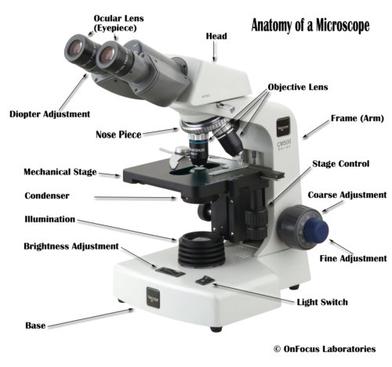
Labeling the Parts of the Microscope | Microscope World Resources Labeling the Parts of the Microscope This activity has been designed for use in homes and schools. Each microscope layout (both blank and the version with answers) are available as PDF downloads. You can view a more in-depth review of each part of the microscope here. Download the Label the Parts of the Microscope PDF printable version here. Compound Microscope- Definition, Labeled Diagram, Principle, Parts, Uses Parts of a Compound Microscope Eyepiece And Body Tube. The eyepiece is the lens through which the viewer looks to see the specimen. It usually contains a 10X or 15X power lens. The body tube connects the eyepiece to the objective lenses. Objectives and Stage Clips Objective Lenses are one of the most important parts of a Compound Microscope. Microscope Parts and Functions Here are the important compound microscope parts... Eyepiece: The lens the viewer looks through to see the specimen. The eyepiece usually contains a 10X or 15X power lens. Diopter Adjustment: Useful as a means to change focus on one eyepiece so as to correct for any difference in vision between your two eyes. Parts of a microscope with functions and labeled diagram - Microbe Notes There are three structural parts of the microscope i.e. head, base, and arm. Head - This is also known as the body. It carries the optical parts in the upper part of the microscope. Base - It acts as microscopes support. It also carries microscopic illuminators.
List: Parts of a Microscope and their Function | Pathwooded
Parts of a Compound Microscope - Labeled (with diagrams) A compound microscope is known as a high-power microscope that enables you to achieve a high level of magnification. Smaller specimens can be thoroughly viewed using a compound microscope. Let us take a look at the different parts of a compound microscope and understand each key component.
1.2: Microscopes - Biology LibreTexts
Compound Microscope Parts - Labeled Diagram and their Functions There are three major structural parts of a compound microscope. The head includes the upper part of the microscope, which houses the most critical optical components, and the eyepiece tube of the microscope. The base acts as the foundation of microscopes and houses the illuminator. The arm connects between the base and the head parts.

Compound Microscope Parts, Functions, and Labeled Diagram ...
Compound Microscope Labeled Diagram | Quizlet Compound Microscope Labeled + − Flashcards Learn Test Match Created by meganplocher734 Terms in this set (14) Eyepiece/Ocular lens Contains the ocular lens Body tube A hollow cylinder that holds the eyepiece. Arm Part that supports the microscope. Stage Supports the slide or specimen Coarse adjustment Knob
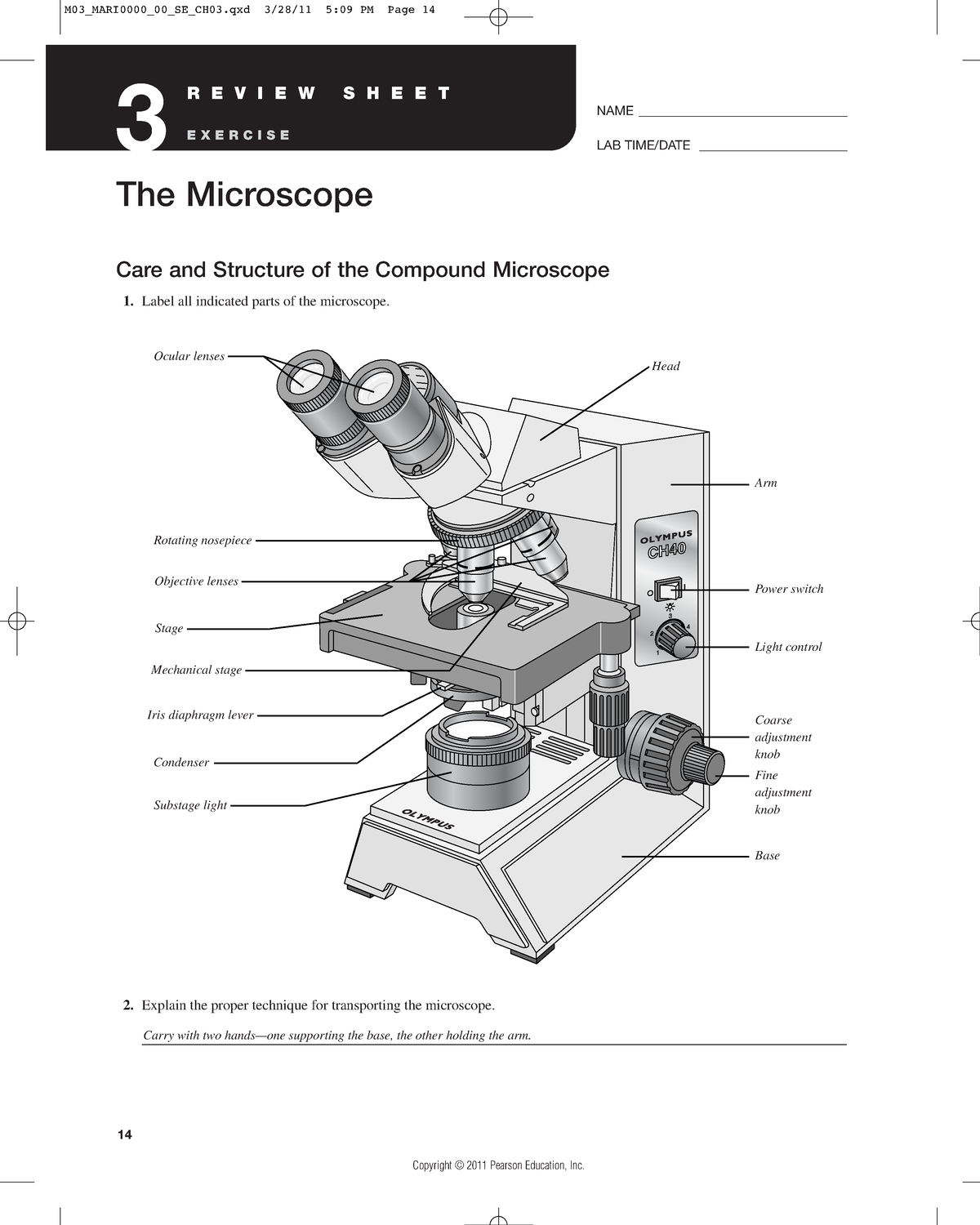
Assignment 3 microscope - Care and Structure of the Compound ...
16 Parts of a Compound Microscope: Diagrams and Video The 16 core parts of a compound microscope are: Head (Body) Arm Base Eyepiece Eyepiece tube Objective lenses Revolving Nosepiece (Turret) Rack stop Coarse adjustment knobs Fine adjustment knobs Stage Stage clips Aperture Illuminator Condenser Diaphragm Video: Parts of a compound Microscope with Diagram Explained
1.5: Microscopy - Biology LibreTexts
Microscopy: Intro to microscopes & how they work (article) - Khan Academy Magnification is a measure of how much larger a microscope (or set of lenses within a microscope) causes an object to appear. For instance, the light microscopes typically used in high schools and colleges magnify up to about 400 times actual size. So, something that was 1 mm wide in real life would be 400 mm wide in the microscope image.

This is a common compound microscope. Label its parts from A ...

This is a common compound microscope. What the labelling D ...
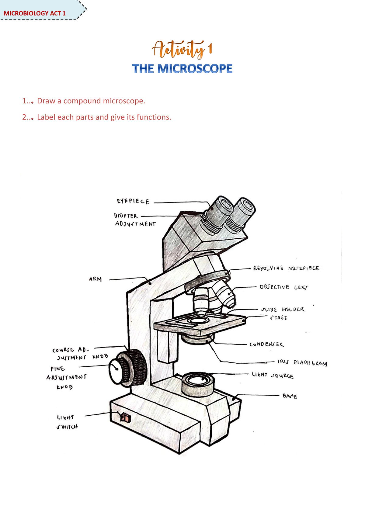
Microscope Activity - MICROBIOLOGY - 1... Draw a compound ...

Microscope Components - Science Quiz - Seterra
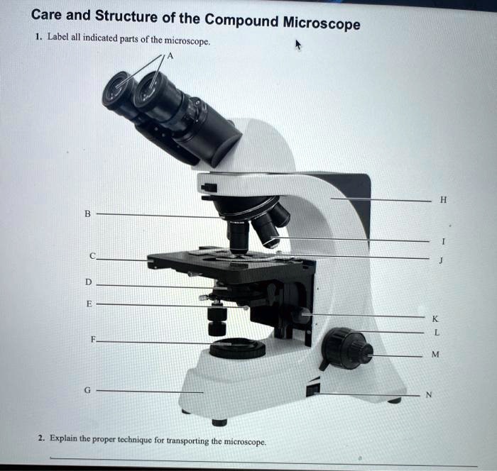
SOLVED: Care and Structure of the Compound Microscope Label ...
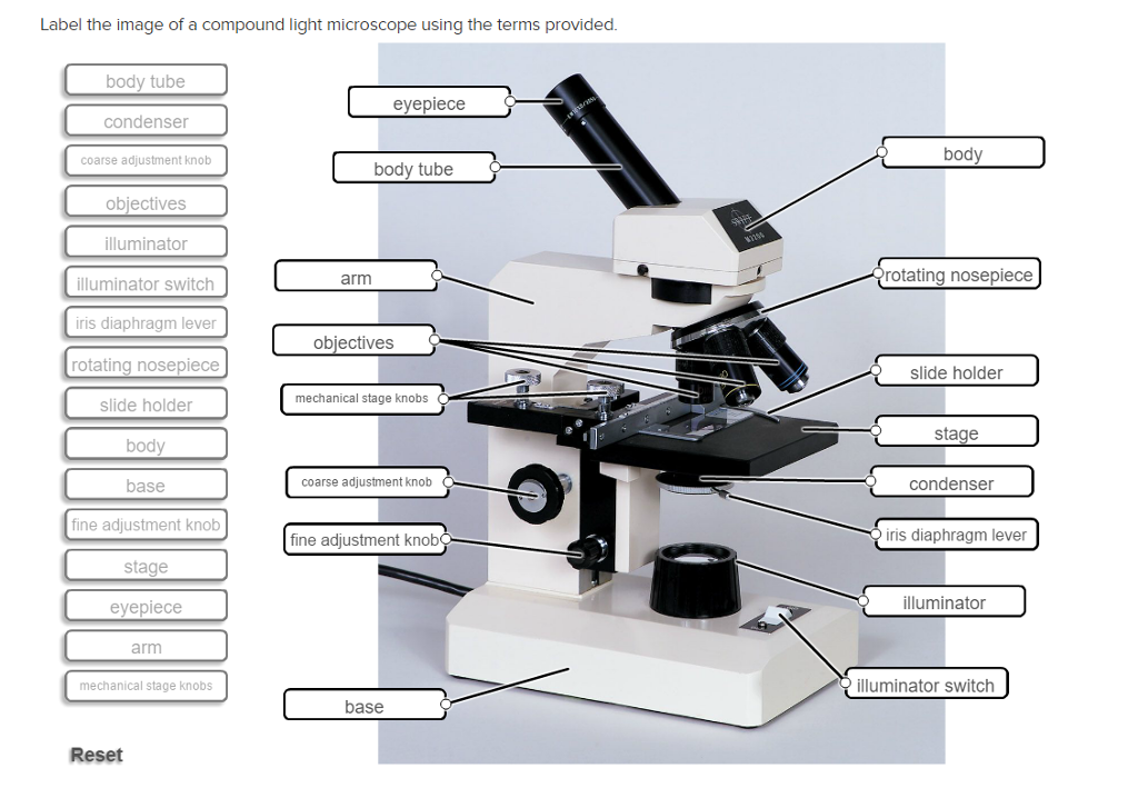
Solved Label the image of a compound light microscope using ...
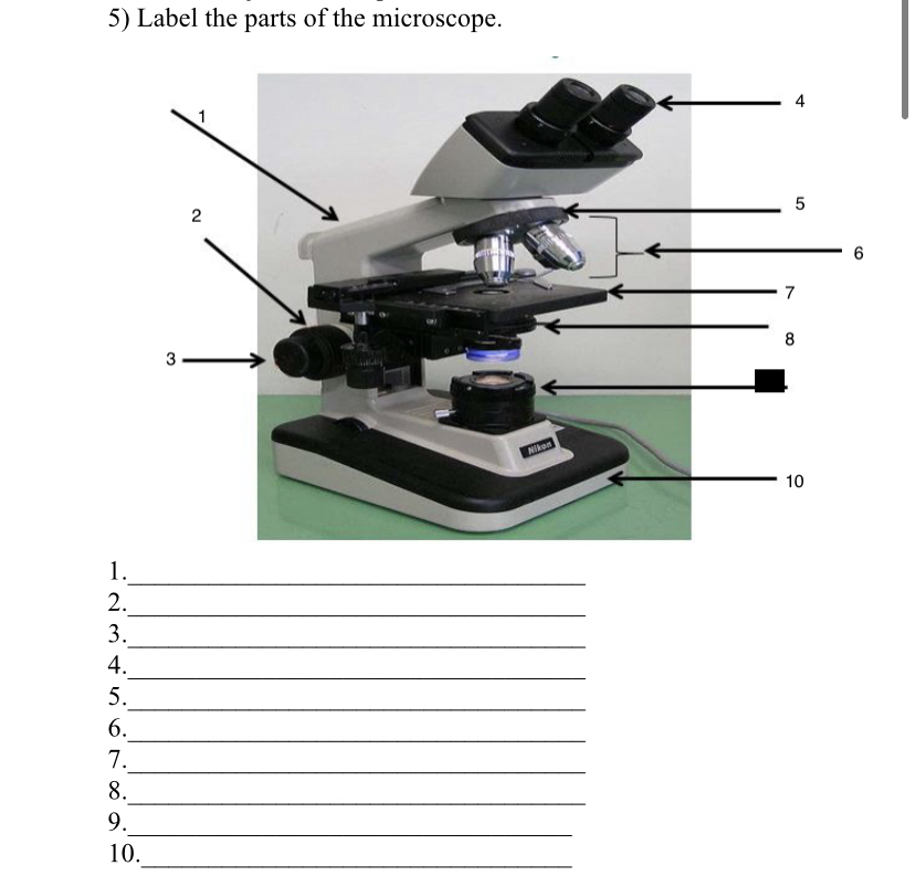
Answered: 5) Label the parts of the microscope. 1… | bartleby

Parts of Stereo Microscope (Dissecting microscope) – labeled ...

Parts of a Microscope - SmartSchool Systems

What are the Parts of a Microscope? - BYJU'S Biology

Microscope Diagram Labeled, Unlabeled and Blank | Parts of a ...

label the parts of the compound microscope - Brainly.ph
Microscope Parts and Functions

Label diagram of compound microscope - Science - The ...
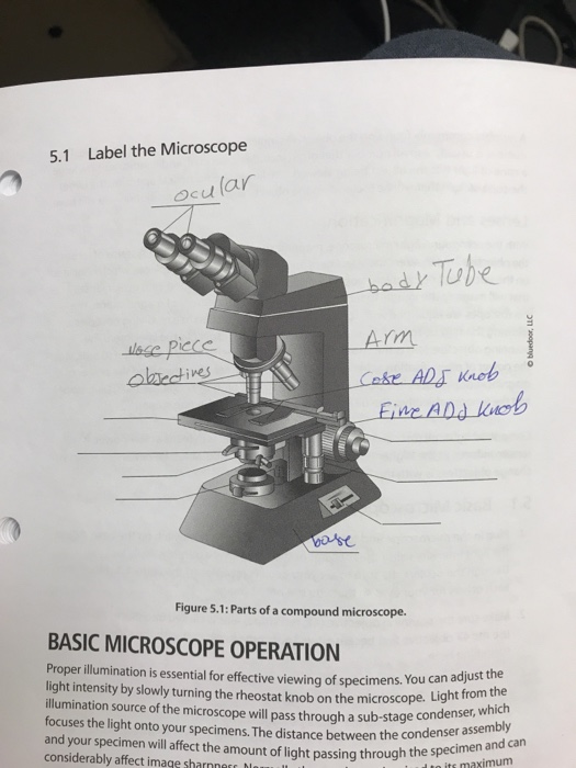
Solved 5.1 Label the Microscope Ocu lar bad Tuhe Arm Lake ...

How to draw compound of Microscope easily - step by step

Label the microscope — Science Learning Hub
Compound Microscope. Label the numbered parts of the ...

II- Label the parts of a Compound Microscope. For numbers ...

Label the parts of a Compound Microscope.docx - A. 1. Label ...
Solved Nikon Parts of the compound microscope Write the ...

This is a common compound microscope. Label its parts from A ...

Pin on Science for kids
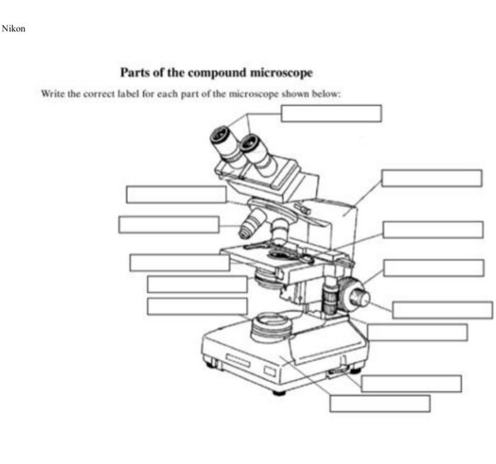
Solved Nikon Parts of the compound microscope Write the ...

Compound Microscope Parts, Function, & Diagram | What is a ...

Compound Microscope Principle, Parts, Diagram Definition ...
Solved) - Care and Structure of the Compound Microscope 1 ...

Parts of the microscope activity

Living Environment Course

Simple Microscope - Diagram (Parts labelled), Principle ...
Microscope Parts & Specifications Labeled Diagram ...

Parts of a Microscope with Their Functions • Microbe Online

Parts Of A Compound Microscope Worksheet
What are the functions of the different parts of a microscope ...

Biology Microscope Labeling and Definitions (Light/Compound ...
Label The Parts Of A Microscope Teaching Resources | TPT

Parts of a Microscope with Their Functions • Microbe Online
Microscope Parts Quiz
Post a Comment for "43 label the parts of a compound microscope"